Department of Biochemistry and Molecular and Cell Biology, University of California San Diego, La Jolla, California, USA.
BMC Neurosci. 2011 Aug 3;12:79. doi: 10.1186/1471-2202-12-79.
α-synuclein [α-Syn]-mediated activation of GSK-3β leading to increases in hyperphosphorylated Tau has been shown by us to occur in striata of Parkinson's diseased [PD] patients and in animal models of PD. In Alzheimer's disease, tauopathy exists in several brain regions; however, the pattern of distribution of tauopathy in other brain regions of PD or in animal models of PD is not known. The current studies were undertaken to analyze the distribution of tauopathy in different brain regions in a widely used mouse model of PD, the α-Syn overexpressing mouse.
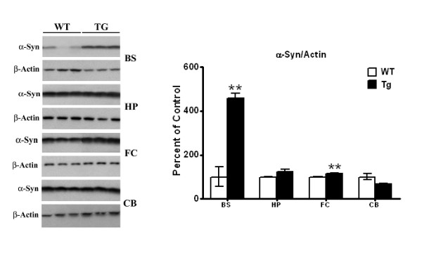
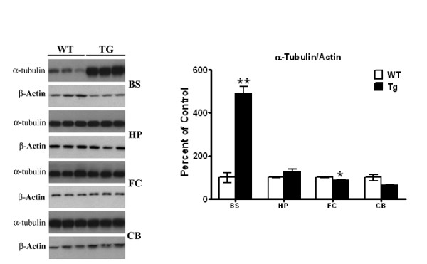
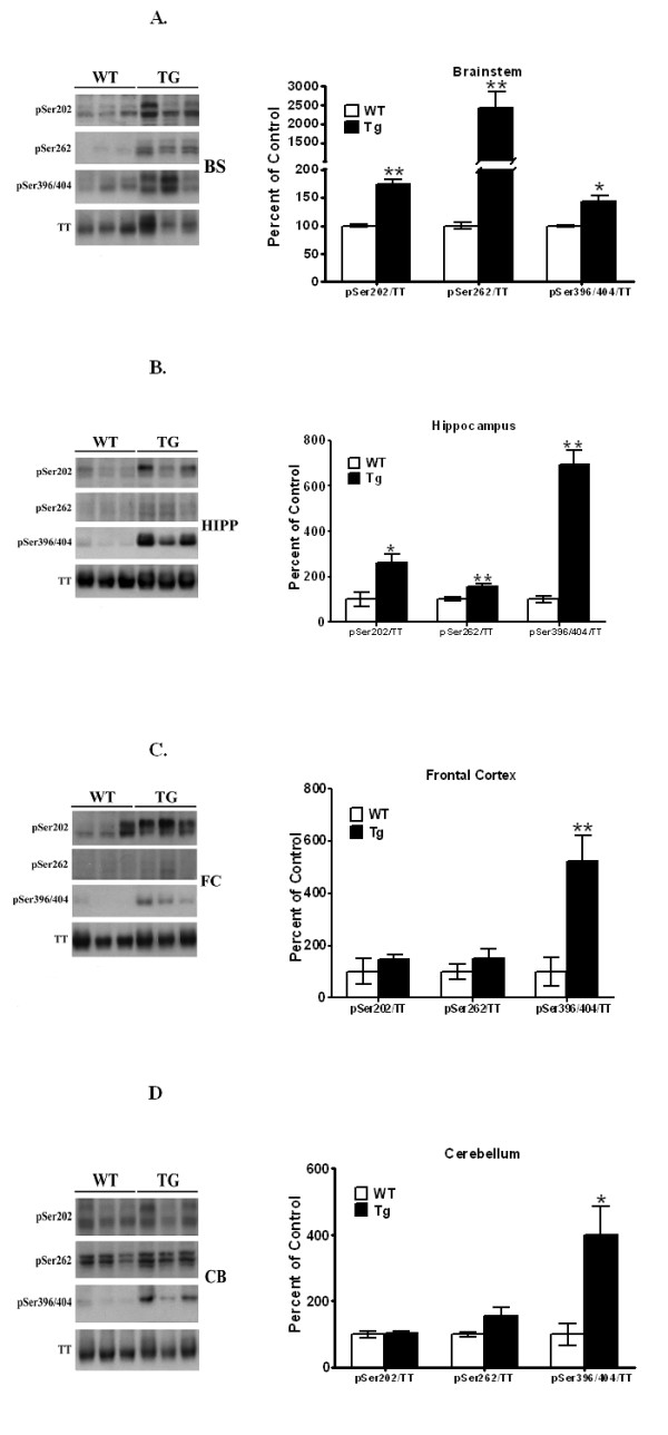
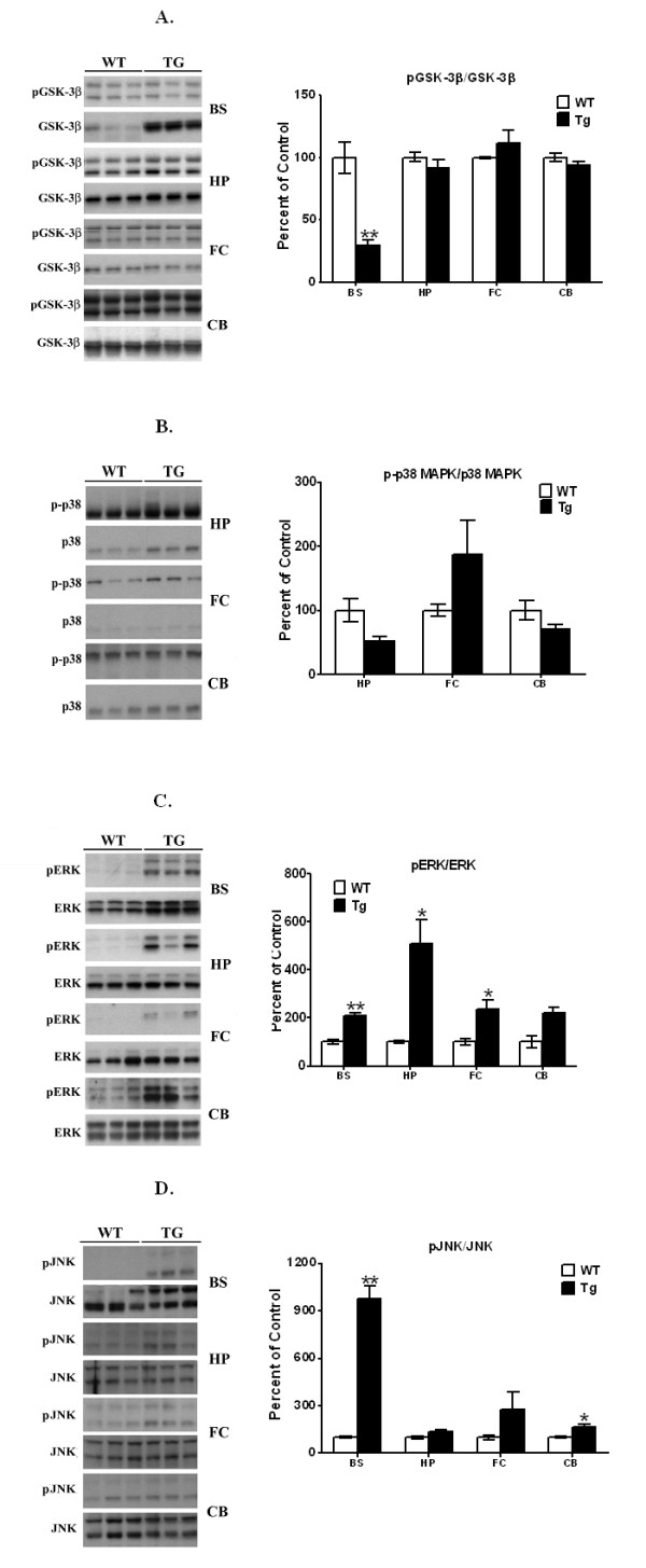
High levels of α-Syn levels were seen in the brain stem, with a much smaller increase in the frontal cortex; neither cerebellum nor hippocampus showed any overexpression of α-Syn. Elevated levels of p-Tau, hyperphosphorylated at Ser202, Ser262 and Ser396/404, were seen in brain stem, with lower levels seen in hippocampus. In both frontal cortex and cerebellum, increases were seen only in p-Ser396/404 Tau, but not in p-Ser202 and p-Ser262. p-GSK-3β levels were not elevated in any of the brain regions, although total GSK-3β was elevated in brain stem. p-p38MAPK levels were unchanged in all brain regions examined, while p-ERK levels were elevated in brain stem, hippocampus and cerebellum, but not the frontal cortex. p-JNK levels were increased in brain stem and cerebellum but not in the frontal cortex or hippocampus. Elevated levels of free tubulin, indicating microtubule destabilization, were seen only in the brain stem.
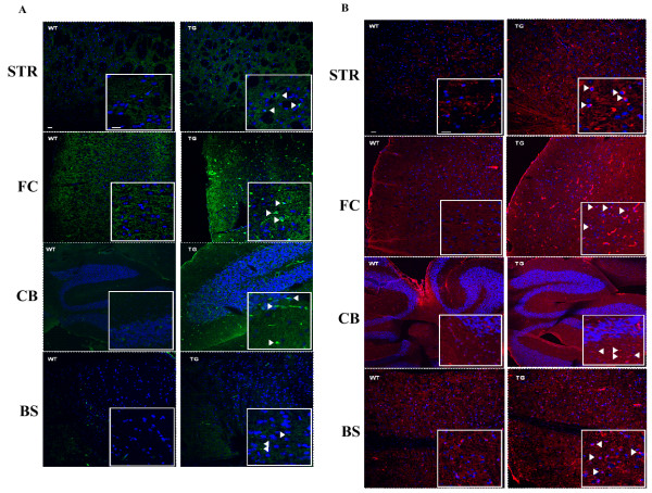
Our combined data suggest that in this animal model of PD, tauopathy, along with microtubule destabilization, exists primarily in the brain stem and striatum, which are also the two major brain regions known to express high levels of α-Syn and undergo the highest levels of degeneration in human PD. Thus, tauopathy in PD may have a very restricted pattern of distribution.
我们已经证明,在帕金森病(PD)患者的纹状体和 PD 动物模型中,α-突触核蛋白[α-Syn]-介导的 GSK-3β 激活导致过度磷酸化 Tau 的增加。在阿尔茨海默病中,tau 病存在于几个大脑区域;然而,在 PD 或 PD 动物模型的其他大脑区域中 tau 病的分布模式尚不清楚。目前的研究旨在分析广泛使用的 PD 小鼠模型,即α-Syn 过表达小鼠中不同大脑区域 tau 病的分布。
在脑干中观察到高水平的 α-Syn,而在额叶皮层中则有较小的增加;小脑和海马体均未表现出任何α-Syn 的过度表达。在脑干中观察到升高的 p-Tau 水平,在 Ser202、Ser262 和 Ser396/404 处过度磷酸化,而在海马体中则观察到较低水平。在前额叶皮层和小脑体中,仅观察到 p-Ser396/404 Tau 的增加,而 p-Ser202 和 p-Ser262 则没有增加。在所有研究的脑区中,p-GSK-3β 水平均未升高,尽管脑干部位的总 GSK-3β 升高。p-p38MAPK 水平在所有检查的脑区中均无变化,而 p-ERK 水平在脑干部、海马体和小脑体中升高,但在前额叶皮层中则没有。p-JNK 水平在前脑干部和小脑体中升高,但在前额叶皮层或海马体中则没有升高。仅在脑干部观察到游离微管蛋白水平升高,表明微管不稳定。
我们的综合数据表明,在这种 PD 动物模型中,tau 病与微管不稳定一起,主要存在于脑干和纹状体中,这也是已知在人类 PD 中表达高水平 α-Syn 并经历最高水平退化的两个主要大脑区域。因此,PD 中的 tau 病可能具有非常受限的分布模式。