• 文献检索
  • 文档翻译
  • 深度研究
  • 学术资讯
  • Suppr Zotero 插件Zotero 插件
  • 邀请有礼
  • 套餐&价格
  • 历史记录
应用&插件
Suppr Zotero 插件Zotero 插件浏览器插件Mac 客户端Windows 客户端微信小程序
定价
高级版会员购买积分包购买API积分包
服务
文献检索文档翻译深度研究API 文档MCP 服务
关于我们
关于 Suppr公司介绍联系我们用户协议隐私条款
关注我们

Suppr 超能文献

核心技术专利:CN118964589B侵权必究
粤ICP备2023148730 号-1Suppr @ 2026

文献检索

告别复杂PubMed语法,用中文像聊天一样搜索,搜遍4000万医学文献。AI智能推荐,让科研检索更轻松。

立即免费搜索

文件翻译

保留排版,准确专业,支持PDF/Word/PPT等文件格式,支持 12+语言互译。

免费翻译文档

深度研究

AI帮你快速写综述,25分钟生成高质量综述,智能提取关键信息,辅助科研写作。

立即免费体验

多连接前沿™冠状动脉分叉支架扩张:人体解剖和猪心样本的微计算机断层扫描评估。

Expansion of the Multi-Link Frontier™ coronary bifurcation stent: micro-computed tomographic assessment in human autopsy and porcine heart samples.

机构信息

I. Department of Medicine, Faculty of Medicine Mannheim, University of Heidelberg, Mannheim, Germany.

出版信息

PLoS One. 2011;6(7):e21778. doi: 10.1371/journal.pone.0021778. Epub 2011 Jul 21.

DOI:10.1371/journal.pone.0021778
PMID:21814552
原文链接:https://pmc.ncbi.nlm.nih.gov/articles/PMC3140974/
Abstract

BACKGROUND

Treatment of coronary bifurcation lesions remains challenging, beyond the introduction of drug eluting stents. Dedicated stent systems are available to improve the technical approach to the treatment of these lesions. However dedicated stent systems have so far not reduced the incidence of stent restenosis. The aim of this study was to assess the expansion of the Multi-Link (ML) Frontier™ stent in human and porcine coronary arteries to provide the cardiologist with useful in-vitro information for stent implantation and selection.

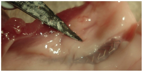
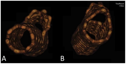
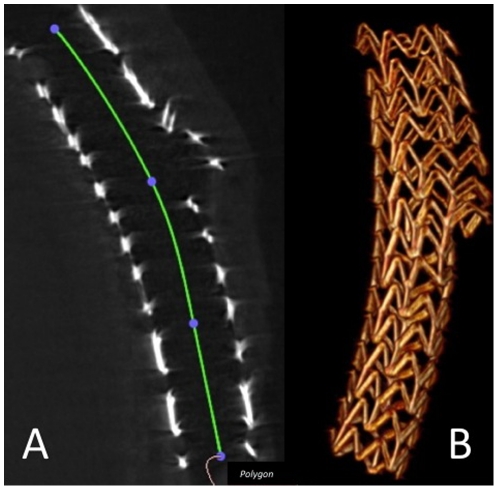
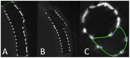
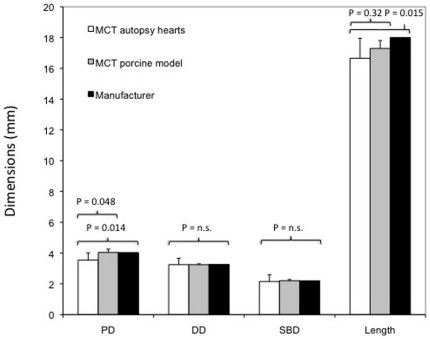
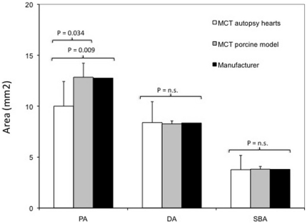
METHODOLOGY/PRINCIPAL FINDINGS: Nine ML Frontier™ stents were implanted in seven human autopsy heart samples with known coronary artery disease and five ML Frontier™ stents were implanted in five porcine hearts. Proximal, distal and side branch diameters (PD, DD, SBD, respectively), corresponding opening areas (PA, DA, SBA) and the mean stent length (L) were assessed by micro-computed tomography (micro-CT). PD and PA were significantly smaller in human autopsy heart samples than in porcine heart samples (3.54±0.47 mm vs. 4.04±0.22 mm, p = 0.048; 10.00±2.42 mm(2) vs. 12.84±1.38 mm(2), p = 0.034, respectively) and than those given by the manufacturer (3.54±0.47 mm vs. 4.03 mm, p = 0.014). L was smaller in human autopsy heart samples than in porcine heart samples, although data did not reach significance (16.66±1.30 mm vs. 17.30±0.51 mm, p = 0.32), and significantly smaller than that given by the manufacturer (16.66±1.30 mm vs. 18 mm, p = 0.015).

CONCLUSIONS/SIGNIFICANCE: Micro-CT is a feasible tool for exact surveying of dedicated stent systems and could make a contribution to the development of these devices. The proximal diameter and proximal area of the stent system were considerably smaller in human autopsy heart samples than in porcine heart samples and than those given by the manufacturer. Special consideration should be given to the stent deployment procedure (and to the follow-up) of dedicated stent systems, considering final intravascular ultrasound or optical coherence tomography to visualize (and if necessary optimize) stent expansion.

摘要

背景

除了药物洗脱支架的应用外,冠状动脉分叉病变的治疗仍然具有挑战性。专用支架系统可用于改善这些病变的治疗技术方法。然而,专用支架系统迄今并未降低支架内再狭窄的发生率。本研究旨在评估 Multi-Link(ML)Frontier™支架在人体和猪冠状动脉中的扩张情况,为心脏介入医生提供支架植入和选择的有用的体外信息。

方法/主要发现:在 7 例已知冠状动脉疾病的人体尸检心脏样本中植入了 9 个 ML Frontier™支架,在 5 例猪心脏中植入了 5 个 ML Frontier™支架。通过微计算机断层扫描(micro-CT)评估近端、远端和侧支直径(PD、DD、SBD,分别)、相应的开口面积(PA、DA、SBA)和平均支架长度(L)。人体尸检心脏样本中的 PD 和 PA 明显小于猪心脏样本(3.54±0.47 mm 比 4.04±0.22 mm,p=0.048;10.00±2.42 mm² 比 12.84±1.38 mm²,p=0.034),也小于制造商提供的数据(3.54±0.47 mm 比 4.03 mm,p=0.014)。人体尸检心脏样本中的 L 小于猪心脏样本,尽管数据没有显著差异(16.66±1.30 mm 比 17.30±0.51 mm,p=0.32),但明显小于制造商提供的数据(16.66±1.30 mm 比 18 mm,p=0.015)。

结论

微计算机断层扫描是一种用于专用支架系统精确测量的可行工具,并可能为这些设备的开发做出贡献。在人体尸检心脏样本中,支架系统的近端直径和近端面积明显小于猪心脏样本和制造商提供的数据。应特别注意专用支架系统的支架展开过程(以及后续过程),考虑进行最终的血管内超声或光学相干断层扫描以可视化(并在必要时优化)支架扩张情况。

https://cdn.ncbi.nlm.nih.gov/pmc/blobs/eb77/3140974/49cae43f48e0/pone.0021778.g006.jpg
https://cdn.ncbi.nlm.nih.gov/pmc/blobs/eb77/3140974/fa7f17762cd7/pone.0021778.g001.jpg
https://cdn.ncbi.nlm.nih.gov/pmc/blobs/eb77/3140974/2ff87ed168ed/pone.0021778.g002.jpg
https://cdn.ncbi.nlm.nih.gov/pmc/blobs/eb77/3140974/e210b53d081c/pone.0021778.g003.jpg
https://cdn.ncbi.nlm.nih.gov/pmc/blobs/eb77/3140974/e853f5ebaa64/pone.0021778.g004.jpg
https://cdn.ncbi.nlm.nih.gov/pmc/blobs/eb77/3140974/ce9576765074/pone.0021778.g005.jpg
https://cdn.ncbi.nlm.nih.gov/pmc/blobs/eb77/3140974/49cae43f48e0/pone.0021778.g006.jpg

相似文献

1
Expansion of the Multi-Link Frontier™ coronary bifurcation stent: micro-computed tomographic assessment in human autopsy and porcine heart samples.多连接前沿™冠状动脉分叉支架扩张:人体解剖和猪心样本的微计算机断层扫描评估。
PLoS One. 2011;6(7):e21778. doi: 10.1371/journal.pone.0021778. Epub 2011 Jul 21.
2
First-in-man study of paclitaxel-eluting stent BiOSS (Bifurcation Optimisation Stent System) dedicated for coronary bifurcation stenoses: three months results.紫杉醇洗脱支架 BiOSS(分叉优化支架系统)治疗冠状动脉分叉病变的首例人体研究:三个月结果。
Kardiol Pol. 2012;70(1):45-52.
3
The Frontier stent registry: safety and feasibility of a novel dedicated stent for the treatment of bifurcation coronary artery lesions.前沿支架注册研究:一种用于治疗冠状动脉分叉病变的新型专用支架的安全性和可行性
J Am Coll Cardiol. 2005 Aug 16;46(4):592-8. doi: 10.1016/j.jacc.2005.05.033.
4
Acute and long-term outcomes of the novel side access (SLK-View) stent for bifurcation coronary lesions: a multicenter nonrandomized feasibility study.新型侧入路(SLK-View)支架治疗冠状动脉分叉病变的急性和长期预后:一项多中心非随机可行性研究
Catheter Cardiovasc Interv. 2006 Feb;67(2):198-206. doi: 10.1002/ccd.20556.
5
Drug-eluting stents in bifurcation lesions: to stent one branch or both?分叉病变中的药物洗脱支架:支架置入一个分支还是两个分支?
Catheter Cardiovasc Interv. 2006 Dec;68(6):891-6. doi: 10.1002/ccd.20870.
6
Healing responses after bifurcation stenting with the dedicated TRYTON Side-Branch Stent™ in combination with XIENCE-V™ stents: a clinical, angiography, fractional flow reserve, and optical coherence tomography study: the PYTON (Prospective evaluation of the TRYTON Side-Branch Stent™ with an additional XIENCE-v™ everolimus-eluting stent in coronary bifurcation lesions) study.采用专用 TRYTON 分支支架™联合 XIENCE-V™支架治疗分叉病变后的愈合反应:一项临床、血管造影、血流储备分数和光学相干断层成像研究:PYTON(前瞻性评价 TRYTON 分支支架™与冠状动脉分叉病变中额外的 XIENCE-v™依维莫司洗脱支架的疗效)研究。
Catheter Cardiovasc Interv. 2013 Feb;81(3):E155-64. doi: 10.1002/ccd.24536. Epub 2012 Nov 14.
7
Analysis of left main coronary artery bifurcation lesions treated with biolimus-eluting DEVAX AXXESS plus nitinol self-expanding stent: intravascular ultrasound results of the AXXENT trial.生物雷帕霉素洗脱DEVAX AXXESS联合镍钛诺自膨胀支架治疗左主干冠状动脉分叉病变的分析:AXXENT试验的血管内超声结果
Catheter Cardiovasc Interv. 2009 Jan 1;73(1):34-41. doi: 10.1002/ccd.21765.
8
Regular Drug-Eluting Stent vs Dedicated Coronary Bifurcation BiOSS Expert Stent: Multicenter Open-Label Randomized Controlled POLBOS I Trial.普通药物洗脱支架与专用冠状动脉分叉BiOSS专家支架对比:多中心开放标签随机对照POLBOS I试验
Can J Cardiol. 2015 May;31(5):671-8. doi: 10.1016/j.cjca.2014.12.024. Epub 2014 Dec 24.
9
Three-dimensional modeling of double-stent techniques at the left main coronary artery bifurcation using micro-focus X-ray computed tomography.使用微焦点X射线计算机断层扫描对左主冠状动脉分叉处双支架技术进行三维建模。
Catheter Cardiovasc Interv. 2007 Aug 1;70(2):211-20. doi: 10.1002/ccd.21133.
10
A modified balloon crush approach improves side branch access and side branch stent apposition during crush stenting of coronary bifurcation lesions.一种改良的球囊挤压方法可改善冠状动脉分叉病变挤压支架置入过程中的边支血管入路及边支支架贴壁情况。
Catheter Cardiovasc Interv. 2006 Sep;68(3):365-71. doi: 10.1002/ccd.20791.

引用本文的文献

1
Assessment of Nit-Occlud atrial septal defect occluder device healing process using micro-computed tomography imaging.应用 micro-computed tomography 成像评估 Nit-Occlud 房间隔缺损封堵器的愈合过程。
PLoS One. 2023 Apr 24;18(4):e0284471. doi: 10.1371/journal.pone.0284471. eCollection 2023.
2
Management of Patients on Non-Vitamin K Antagonist Oral Anticoagulants in the Acute Care and Periprocedural Setting: A Scientific Statement From the American Heart Association.急性护理和围手术期非维生素K拮抗剂口服抗凝剂治疗患者的管理:美国心脏协会科学声明
Circulation. 2017 Mar 7;135(10):e604-e633. doi: 10.1161/CIR.0000000000000477. Epub 2017 Feb 6.
3

本文引用的文献

1
Micro-computed tomographic assessment following extremely oversized partial postdilatation of drug-eluting stents.药物洗脱支架超大号后扩张后行微计算机断层扫描评估。
EuroIntervention. 2010 May;6(1):141-8.
2
Current preservation technology and future prospects of thoracic organs. Part 2: heart.胸器官的当前保存技术和未来前景。第 2 部分:心脏。
Curr Opin Organ Transplant. 2010 Apr;15(2):156-9. doi: 10.1097/MOT.0b013e328337343f.
3
Randomized comparison of coronary bifurcation stenting with the crush versus the culotte technique using sirolimus eluting stents: the Nordic stent technique study.
Evaluation of occurring complications after flow diverter treatment of elastase-induced aneurysm in rabbits using micro-CT and MRI at 9.4 T.
使用9.4T微型计算机断层扫描(micro-CT)和磁共振成像(MRI)对兔弹性蛋白酶诱导的动脉瘤进行血流导向治疗后发生的并发症的评估。
Neuroradiology. 2016 Oct;58(10):987-996. doi: 10.1007/s00234-016-1730-x. Epub 2016 Jul 25.
4
Comparison of high-resolution X-ray and micro-CT for experimental evaluation of intracranial stent prototypes: quality evaluation beyond CE mark.高分辨率X射线与微型计算机断层扫描用于颅内支架原型实验评估的比较:超越CE标志的质量评估
Neuroradiology. 2014 Apr;56(4):315-23. doi: 10.1007/s00234-014-1324-4. Epub 2014 Jan 31.
5
Compound ex vivo and in silico method for hemodynamic analysis of stented arteries.载药支架血管血流动力学的复合离体与计算方法研究。
PLoS One. 2013;8(3):e58147. doi: 10.1371/journal.pone.0058147. Epub 2013 Mar 13.
随机比较使用西罗莫司洗脱支架的分叉病变支架置入术的 crush 与 culotte 技术:北欧支架技术研究。
Circ Cardiovasc Interv. 2009 Feb;2(1):27-34. doi: 10.1161/CIRCINTERVENTIONS.108.804658. Epub 2009 Feb 10.
4
Percutaneous coronary intervention of bifurcation lesions: state-of-the-art. Insights from the second meeting of the European Bifurcation Club.分叉病变的经皮冠状动脉介入治疗:最新进展。来自欧洲分叉病变俱乐部第二次会议的见解。
EuroIntervention. 2007 May;3(1):44-9.
5
Very late stent thrombosis of sirolimus-eluting stent combined with late stent malapposition and aneurysm formation.西罗莫司洗脱支架的极晚期支架内血栓形成合并晚期支架贴壁不良和动脉瘤形成。
J Invasive Cardiol. 2009 Sep;21(9):E175-7.
6
Predictors and prognosis of stent fracture after sirolimus-eluting stent implantation.西罗莫司洗脱支架置入后支架断裂的预测因子和预后。
Circ J. 2009 Nov;73(11):2036-41. doi: 10.1253/circj.cj-09-0343. Epub 2009 Aug 28.
7
Percutaneous coronary intervention for bifurcation lesions: 2008 consensus document from the fourth meeting of the European Bifurcation Club.冠状动脉分叉病变的经皮冠状动脉介入治疗:欧洲分叉病变俱乐部第四次会议2008年共识文件
EuroIntervention. 2009 May;5(1):39-49. doi: 10.4244/eijv5i1a8.
8
Drug-eluting stents: careful deployment required.药物洗脱支架:需要谨慎植入。
JACC Cardiovasc Interv. 2009 May;2(5):435-6. doi: 10.1016/j.jcin.2009.02.012.
9
A volumetric intravascular ultrasound comparison of early drug-eluting stent thrombosis versus restenosis.药物洗脱支架早期血栓形成与再狭窄的血管内超声体积学比较。
JACC Cardiovasc Interv. 2009 May;2(5):428-34. doi: 10.1016/j.jcin.2009.01.011.
10
Update on dedicated bifurcation stents.专用分叉支架的最新进展。
J Interv Cardiol. 2009 Apr;22(2):150-5. doi: 10.1111/j.1540-8183.2009.00444.x.