Engelen Marc, Westhoff Dunja, de Gans Jan, Nederkoorn Paul J
Department of Neurology, H2,216, Academic Medical Center, University of Amsterdam, PO Box 22660, 1100 DD Amsterdam, The Netherlands.
J Med Case Rep. 2011 Aug 9;5:357. doi: 10.1186/1752-1947-5-357.
Magnetic resonance imaging of the brain in patients with corticobasal degeneration typically shows focal or asymmetric atrophy, usually maximal in the frontoparietal cortex. Many patients who are diagnosed with corticobasal degeneration using current diagnostic criteria do not have classical corticobasal degeneration pathology. Our case is remarkable for the fact that the symptoms and the characteristic magnetic resonance imaging appearance were typical for corticobasal degeneration. However, we were quite convinced that the clinical picture had a vascular etiology. Only a few cases have been reported where the presumed cause for the corticobasal syndrome was multiple brain infarctions bilaterally.
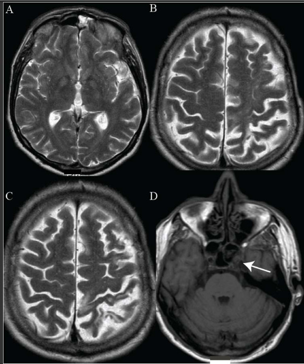
A 64-year-old Caucasian man visited a neurologist because of profound asymmetric sensory and motor disturbances. A magnetic resonance imaging scan of his brain revealed occlusion of his internal carotid artery on the left side with multiple vascular lesions in his left hemisphere and notable atrophy of mainly the left parietal and frontal cortex.
We describe a patient with corticobasal syndrome caused by multiple infarctions, probably caused by emboli of the carotid stenosis. This patient illustrates the fact that the word 'syndrome' should be preferred above 'degeneration' in the name of this disease.
皮质基底节变性患者的脑部磁共振成像通常显示局灶性或不对称性萎缩,通常在额顶叶皮质最为明显。许多根据当前诊断标准被诊断为皮质基底节变性的患者并没有典型的皮质基底节变性病理学表现。我们的病例值得注意的是,其症状和典型的磁共振成像表现符合皮质基底节变性。然而,我们坚信临床症状有血管源性病因。仅有少数病例报道双侧多发性脑梗死被认为是皮质基底节综合征的病因。
一名64岁的白人男性因严重的不对称感觉和运动障碍就诊于神经科医生。他的脑部磁共振成像扫描显示左侧颈内动脉闭塞,左侧半球有多个血管病变,主要是左侧顶叶和额叶皮质明显萎缩。
我们描述了一名由多发性梗死引起的皮质基底节综合征患者,可能是由颈动脉狭窄的栓子所致。该患者表明在这种疾病的命名中,“综合征”一词应优先于“变性”。