Molecular Function & Imaging Program, National Cardiac PET Centre, University of Ottawa Heart Institute, 40 Ruskin Street, Ottawa, K1Y 4W7, Canada.
Cardiovasc Diabetol. 2011 Aug 10;10:75. doi: 10.1186/1475-2840-10-75.
Diabetes mellitus is strongly associated with cardiovascular dysfunction, derived in part from impairment of sympathetic nervous system signaling. Glucose, insulin, and non-esterified fatty acids are potent stimulants of sympathetic activity and norepinephrine (NE) release. We hypothesized that sustained hyperglycemia in the high fat diet-fed streptozotocin (STZ) rat model of sustained hyperglycemia with insulin resistance would exhibit progressive sympathetic nervous dysfunction in parallel with deteriorating myocardial systolic and/or diastolic function.
Cardiac sympathetic nervous integrity was investigated in vivo via biodistribution of the positron emission tomography radiotracer and NE analogue [11C]meta-hydroxyephedrine ([11C]HED). Cardiac systolic and diastolic function was evaluated by echocardiography. Plasma and cardiac NE levels and NE reuptake transporter (NET) expression were evaluated as correlative measurements.
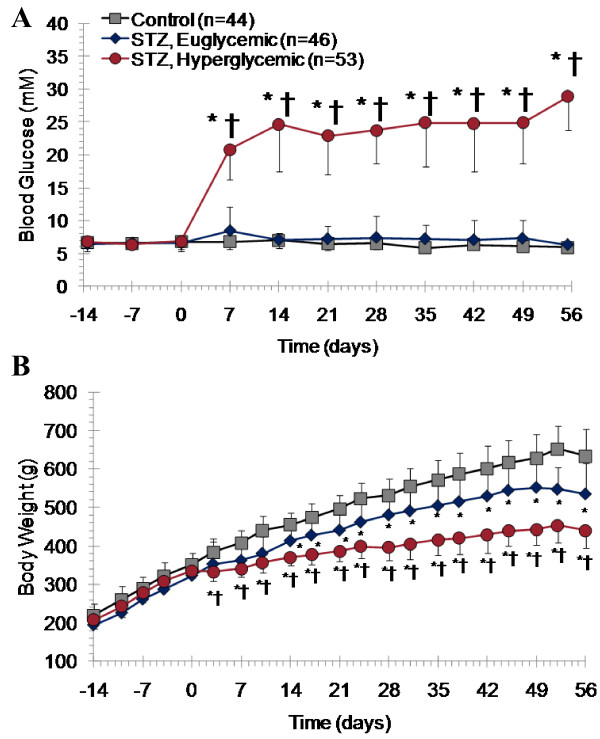
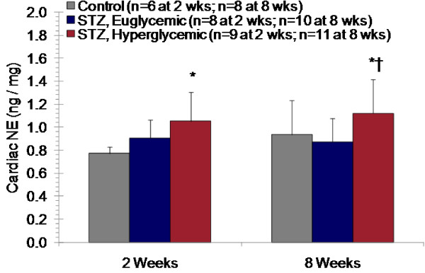
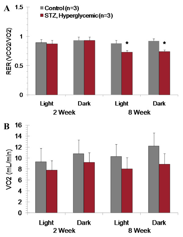
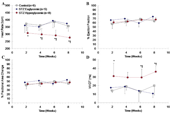
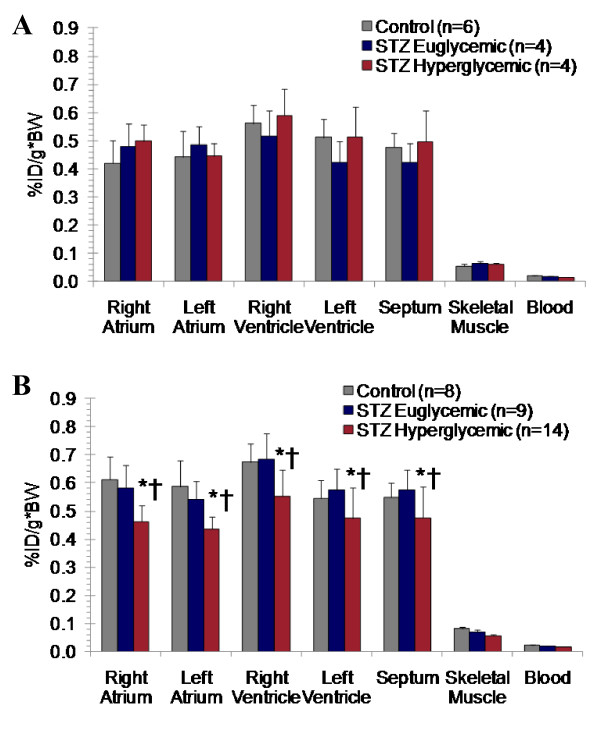
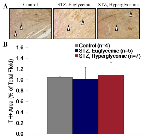
The animal model displays insulin resistance, sustained hyperglycemia, and progressive hypoinsulinemia. After 8 weeks of persistent hyperglycemia, there was a significant 13-25% reduction in [11C]HED retention in myocardium of STZ-treated hyperglycemic but not euglycemic rats as compared to controls. There was a parallel 17% reduction in immunoblot density for NE reuptake transporter, a 1.2 fold and 2.5 fold elevation of cardiac and plasma NE respectively, and no change in sympathetic nerve density. No change in ejection fraction or fractional area change was detected by echocardiography. Reduced heart rate, prolonged mitral valve deceleration time, and elevated transmitral early to atrial flow velocity ratio measured by pulse-wave Doppler in hyperglycemic rats suggest diastolic impairment of the left ventricle.
Taken together, these data suggest that sustained hyperglycemia is associated with elevated myocardial NE content and dysregulation of sympathetic nervous system signaling in the absence of systolic impairment.
糖尿病与心血管功能障碍密切相关,部分原因是交感神经系统信号受损。葡萄糖、胰岛素和非酯化脂肪酸是强烈刺激交感活性和去甲肾上腺素(NE)释放的物质。我们假设,在高血糖饮食喂养的链脲佐菌素(STZ)大鼠模型中,持续的高血糖伴胰岛素抵抗会表现出进行性的交感神经功能障碍,同时伴有心肌收缩和/或舒张功能恶化。
通过正电子发射断层扫描示踪剂和 NE 类似物[11C]meta-羟基苯丙胺([11C]HED)的生物分布,在体内研究心脏交感神经完整性。通过超声心动图评估心肌收缩和舒张功能。评估血浆和心脏去甲肾上腺素水平以及去甲肾上腺素再摄取转运体(NET)表达作为相关测量。
该动物模型表现出胰岛素抵抗、持续高血糖和进行性低胰岛素血症。在持续高血糖 8 周后,与对照组相比,STZ 处理的高血糖但非高血糖大鼠的心肌[11C]HED 保留显著减少 13-25%。NE 再摄取转运体的免疫印迹密度平行降低 17%,心脏和血浆去甲肾上腺素分别升高 1.2 倍和 2.5 倍,而交感神经密度无变化。超声心动图未检测到射血分数或分数面积变化的变化。在高血糖大鼠中,通过脉搏波多普勒测量的心率降低、二尖瓣减速时间延长和二尖瓣早期到心房血流速度比升高,提示左心室舒张功能受损。
综上所述,这些数据表明,持续高血糖与心肌 NE 含量升高和交感神经系统信号调节失调有关,而无收缩功能损害。