Department of Pediatrics, Skåne University Hospital, Lund University, Sweden.
Pediatr Rheumatol Online J. 2011 Aug 11;9(1):22. doi: 10.1186/1546-0096-9-22.
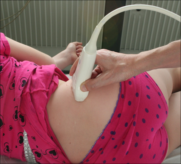
The presence of enthesitis (insertional inflammation) in patients with juvenile idiopathic arthritis (JIA) is difficult to establish clinically and may influence classification and treatment of the disease. We used ultrasonography (US) and color Doppler (CD) imaging to detect enthesitis at the small and deep-seated proximal insertion of the gluteus medius fascia on the posterior iliac crest where clinical diagnosis is difficult. The findings in JIA patients were compared with those obtained in healthy controls and with the patients' MRI results.
Seventy-six proximal gluteus medius insertions were studied clinically (tenderness to palpation of the posterior iliac crest) and by US and CD (echogenicity, thickness, hyperemia) in 38 patients with JIA and in 38 healthy controls, respectively (median age 13 years, range 7-18 years). In addition, an additional MRI examination of the sacroiliac joints and iliac crests was performed in all patients.
In patients with focal, palpable tenderness, US detected decreased echogenicity of the entheses in 53% of the iliac crests (bilateral in 37% and unilateral in 32%). US also revealed significantly thicker entheses in JIA patients compared to healthy controls (p < 0.003 left side, p < 0.001 right side). There was no significant difference in thickness between the left and right sides in individual subjects. Hyperemia was detected by CD in 37% (28/76) of the iliac crests and by contrast-enhanced MRI in 12% (6/50).
According to US, the gluteus medius insertion was thicker in JIA patients than in controls, and it was hypoechoic (enthesitis) in about half of the patients. These findings may represent chronic, inactive disease in some of the patients, because there was only limited Doppler flow and MRI contrast enhancement. The present study indicates that US can be useful as an adjunct to clinical examination for improved assessment of enthesitis in JIA. This may influence disease classification, ambition to treat, and choice of treatment regimen.
在幼年特发性关节炎(JIA)患者中,附着点炎(插入炎症)的存在临床上难以确定,可能影响疾病的分类和治疗。我们使用超声(US)和彩色多普勒(CD)成像来检测臀部中肌筋膜在髂后嵴后部小而深部的插入处的附着点炎,因为临床上难以诊断。将 JIA 患者的发现与健康对照组和患者的 MRI 结果进行比较。
分别对 38 例 JIA 患者和 38 例健康对照者的 76 个近端臀中肌附着处进行了临床(髂后嵴触诊压痛)和 US 和 CD(回声、厚度、充血)研究(中位年龄 13 岁,范围 7-18 岁)。此外,所有患者均进行了额外的骶髂关节和髂嵴 MRI 检查。
在有局灶性、可触及压痛的患者中,US 检测到 53%的髂嵴附着处回声降低(双侧 37%,单侧 32%)。与健康对照组相比,JIA 患者的附着处明显增厚(左侧 p < 0.003,右侧 p < 0.001)。在个别患者中,左右两侧的厚度无显著差异。CD 检测到 37%(28/76)的髂嵴充血,对比增强 MRI 检测到 12%(6/50)。
根据 US,JIA 患者的臀中肌附着处比对照组更厚,约一半的患者回声降低(附着点炎)。这些发现可能代表一些患者的慢性、非活动性疾病,因为只有有限的多普勒血流和 MRI 增强。本研究表明,US 可作为临床检查的辅助手段,用于改善 JIA 附着点炎的评估。这可能影响疾病分类、治疗目标和治疗方案的选择。