Department of Pediatrics, Skåne University Hospital, Lund University, Lund, Sweden.
Pediatr Rheumatol Online J. 2012 Aug 16;10(1):23. doi: 10.1186/1546-0096-10-23.
In juvenile idiopathic arthritis (JIA), the trend towards early therapeutic intervention and the development of new highly effective treatments have increased the need for sensitive and specific imaging. Numerous studies have demonstrated the important role of MRI and US in adult rheumatology. However, investigations of imaging in JIA are rare, and no previous study has been comparing MRI with Doppler ultrasonography (US) for assessment of arthritis. The aim of the present study was to compare the two imaging methods regarding their usefulness for evaluating disease activity in JIA, and to compare the results with those obtained in healthy controls.
In 10 JIA patients (median age 14 years, range 11-18), 11 joints (six wrists, three knees, two ankles) with arthritis were assessed by color Doppler US and MRI. The same imaging modalities were used to evaluate eight joints (three wrists, three knees, two ankles) in six healthy age- and sex-matched controls. The US examinations of both the patients and controls were compared with the MRI findings.
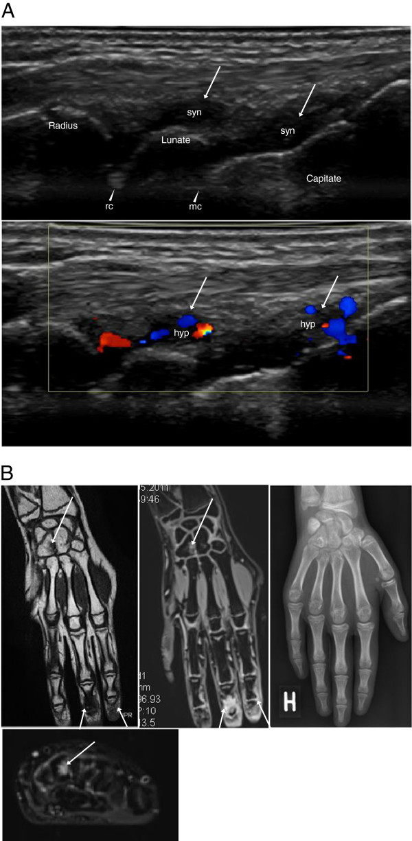
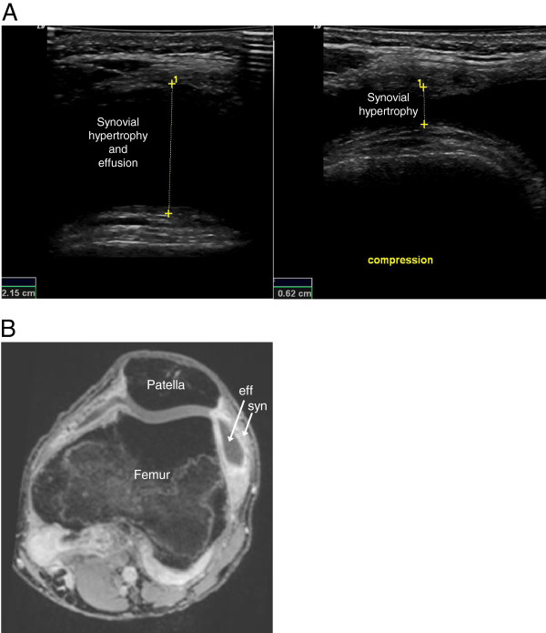
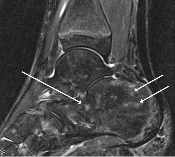
In 10 JIA patients, US detected synovial hypertrophy in 22 areas of 11 joints, 86% of which had synovial hyperemia, and MRI revealed synovitis in 36 areas of the same 11 joints. Erosions were identified by US in two areas of two joints and by MRI in six areas of four joints. Effusion was shown by US in nine areas of six joints and by MRI in 17 areas of five joints. MRI detected juxta-articular bone marrow edema in 16 areas of eight joints.
The results of this pilot study indicate that both MRI and US provide valuable imaging information on disease activity in JIA. Importantly, the two techniques seem to complement each other and give partly different information. Although MRI is considered to be the reference standard for advanced imaging in adult rheumatology, US seems to provide useful imaging information that could make it an option in daily clinical practice, in JIA as well as in adult rheumatology. However, the current work represents a pilot study, and thus our results need to be confirmed in a larger prospective clinical investigation.
在幼年特发性关节炎(JIA)中,早期治疗干预的趋势和新的高效治疗方法的发展增加了对敏感和特异成像的需求。许多研究已经证明了 MRI 和 US 在成人风湿病学中的重要作用。然而,JIA 的影像学研究很少,以前没有研究比较过 MRI 与多普勒超声(US)在关节炎评估中的作用。本研究的目的是比较两种成像方法在评估 JIA 疾病活动中的有用性,并将结果与健康对照组进行比较。
在 10 例 JIA 患者(中位年龄 14 岁,范围 11-18 岁)中,11 个关节(6 个腕关节、3 个膝关节、2 个踝关节)进行彩色多普勒 US 和 MRI 评估。使用相同的成像方式评估 6 名年龄和性别匹配的健康对照组的 8 个关节(3 个腕关节、3 个膝关节、2 个踝关节)。将患者和对照组的 US 检查结果与 MRI 结果进行比较。
在 10 例 JIA 患者中,US 检测到 11 个关节的 22 个部位的滑膜增生,其中 86%的部位有滑膜充血,MRI 显示 11 个关节的 36 个部位有滑膜炎。US 在 2 个关节的 2 个部位和 MRI 在 4 个关节的 6 个部位发现了侵蚀。US 在 6 个关节的 9 个部位和 MRI 在 5 个关节的 17 个部位显示了积液。MRI 在 8 个关节的 16 个部位检测到关节旁骨髓水肿。
这项初步研究的结果表明,MRI 和 US 均可为 JIA 的疾病活动提供有价值的影像学信息。重要的是,这两种技术似乎相互补充,提供了部分不同的信息。尽管 MRI 被认为是成人风湿病学中高级成像的参考标准,但 US 似乎提供了有用的影像学信息,使其成为日常临床实践中的一种选择,不仅在 JIA 中,而且在成人风湿病学中也是如此。然而,目前的工作只是一项初步研究,因此我们的结果需要在更大的前瞻性临床研究中得到证实。