Research Service, Veterans Affairs San Diego Healthcare System San Diego, CA, USA.
Front Integr Neurosci. 2011 Jul 28;5:32. doi: 10.3389/fnint.2011.00032. eCollection 2011.
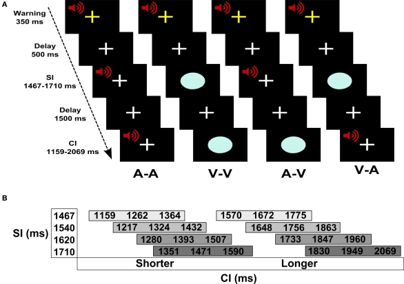
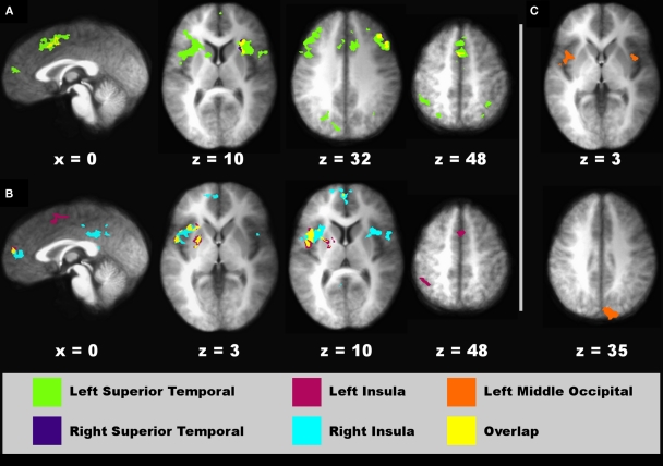
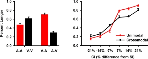
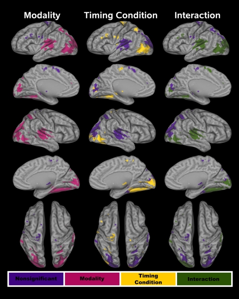
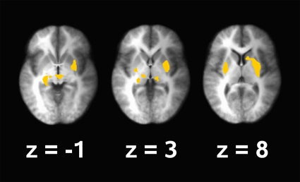
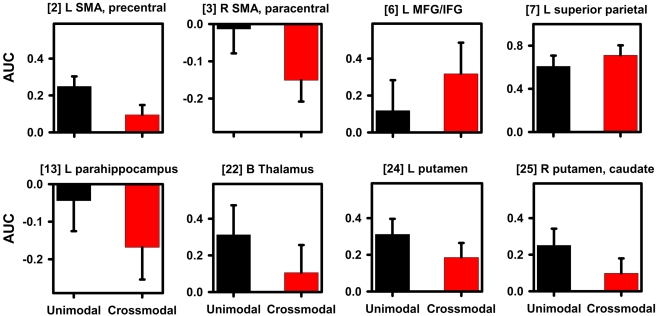
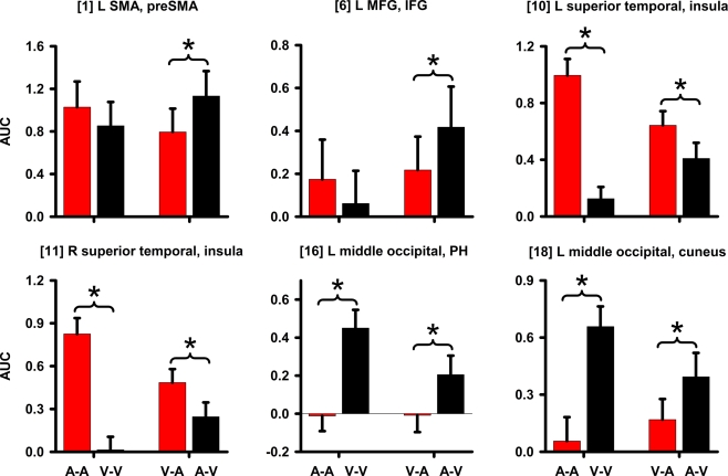
Auditory signals (A) are perceived as lasting longer than visual signals (V) of the same physical duration when they are compared together. Despite considerable debate about how this illusion arises psychologically, the neural underpinnings have not been studied. We used functional magnetic resonance imaging (fMRI) to investigate the neural bases of audiovisual temporal distortions and more generally, intersensory timing. Adults underwent fMRI while judging the relative duration of successively presented standard interval-comparison interval (CI) pairs, which were unimodal (A-A, V-V) or crossmodal (V-A, A-V). Mechanisms of time dilation and compression were identified by comparing the two crossmodal pairs. Mechanisms of intersensory timing were identified by comparing the unimodal and crossmodal conditions. The behavioral results showed that auditory CIs were perceived as lasting longer than visual CIs. There were three novel fMRI results. First, time dilation and compression were distinguished by differential activation of higher-sensory areas (superior temporal, posterior insula, middle occipital), which typically showed stronger effective connectivity when time was dilated (V-A). Second, when time was compressed (A-V) activation was greater in frontal cognitive-control centers, which guide decision making. These areas did not exhibit effective connectivity. Third, intrasensory timing was distinguished from intersensory timing partly by decreased striatal and increased superior parietal activation. These regions showed stronger connectivity with visual, memory, and cognitive-control centers during intersensory timing. Altogether, the results indicate that time dilation and compression arise from the connectivity strength of higher-sensory systems with other areas. Conversely, more extensive network interactions are needed with core timing (striatum) and attention (superior parietal) centers to integrate time codes for intersensory signals.
当听觉信号 (A) 与相同物理时长的视觉信号 (V) 进行比较时,人们会觉得听觉信号持续的时间更长。尽管对于这种错觉是如何在心理上产生的存在大量争议,但尚未研究其神经基础。我们使用功能磁共振成像 (fMRI) 来研究视听时间扭曲的神经基础,更广泛地说,是研究感官间定时。成年人在进行 fMRI 扫描的同时判断相继呈现的标准间隔比较间隔 (CI) 对的相对持续时间,这些间隔对是单模态 (A-A、V-V) 或跨模态 (V-A、A-V)。通过比较两种跨模态对来确定时间扩张和压缩的机制。通过比较单模态和跨模态条件来确定感官间定时的机制。行为结果表明,听觉 CI 被感知为比视觉 CI 持续时间更长。有三个新的 fMRI 结果。首先,通过更高感官区域(颞上区、后岛叶、中枕叶)的差异激活来区分时间扩张和压缩,当时间扩张时(V-A),这些区域通常表现出更强的有效连接。其次,当时间压缩时(A-V),激活更多地出现在额叶认知控制中心,这些中心指导决策。这些区域没有表现出有效连接。第三,通过纹状体和顶叶上回的激活减少以及增强来区分内感官定时和感官间定时。这些区域在感官间定时期间与视觉、记忆和认知控制中心显示出更强的连接。总之,这些结果表明,时间扩张和压缩是由较高感官系统与其他区域的连接强度引起的。相反,需要更广泛的网络相互作用,以与核心定时(纹状体)和注意(顶叶上回)中心整合感官间信号的时间码。