Pablo Luis E, Garcia-Martin Elena, Gazulla Jose, Larrosa Jose M, Ferreras Antonio, Santorelli Filippo M, Benavente Isabel, Vela Ana, Marin Miguel A
Ophthalmology Department, Miguel Servet University Hospital, Zaragoza, Spain.
Mol Vis. 2011;17:1871-6. Epub 2011 Jul 13.
To present full ophthalmologic examination and retinal nerve fiber layer (RNFL) photographs of autosomal recessive spastic ataxia of Charlevoix-Saguenay (ARSACS) patients showing significant increases in RNFL thickness compared to healthy subjects, but without myelinated retinal fibers.
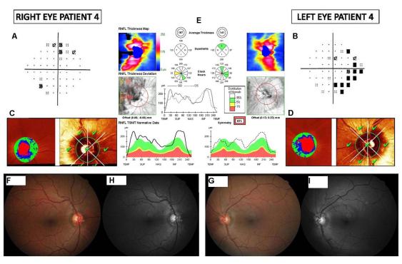
The study design was observational case series. Ten eyes of five patients with molecular confirmation of ARSACS underwent a full ophthalmologic examination that included clinical history, visual acuity, biomicroscopy of the anterior segment, gonioscopy, Goldmann applanation tonometry, central corneal ultrasonic pachymetry, ophthalmoscopy of the posterior segment, standard automatic perimetry (Humphrey field), simultaneous stereophotographs of the optic disc after mydriasis, a series of five red-free digital fundus photographs for RNFL evaluation, topographic analysis of the optic disc using the Heidelberg retina tomography, and measurement of peripapillary RNFL thickness with Cirrus optical coherence tomography.
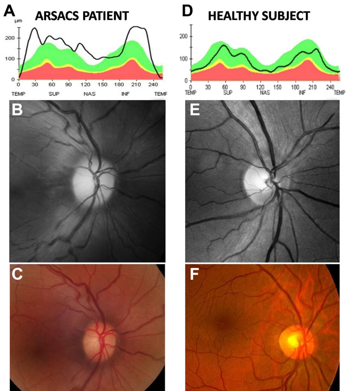
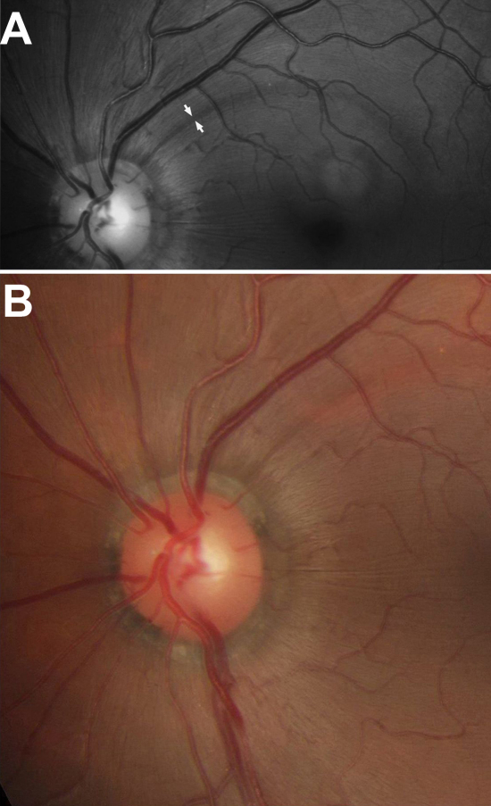
All patients showed abnormal visual fields, normal optic discs with a mild to strikingly increased visibility of RNFL in color stereophotographs, normal Heidelberg tomography, and moderate to markedly increased RNFL thickness in Cirrus tomography (average thickness ranging from 119 μm to 220 μm).
We found evidence of RNFL hypertrophy in ARSACS patients that may have been interpreted as hypermyelinated retinal fibers in previous reports. A revision of ARSACS diagnostic criteria, particularly with regard to retinal alterations, is necessary.
展示夏尔沃 - 萨格奈常染色体隐性痉挛性共济失调(ARSACS)患者的全面眼科检查及视网膜神经纤维层(RNFL)照片,这些患者与健康受试者相比,RNFL厚度显著增加,但无髓鞘化视网膜纤维。
研究设计为观察性病例系列。对5例经分子确诊的ARSACS患者的10只眼睛进行了全面眼科检查,包括临床病史、视力、前段生物显微镜检查、前房角镜检查、Goldmann压平眼压测量、中央角膜超声测厚、后段检眼镜检查、标准自动视野计检查(Humphrey视野)、散瞳后视盘同步立体照片、用于RNFL评估的一系列五张无赤数字眼底照片、使用海德堡视网膜断层扫描对视盘进行地形分析以及用Cirrus光学相干断层扫描测量视乳头周围RNFL厚度。
所有患者均显示视野异常,视盘正常,彩色立体照片中RNFL可见度轻度至显著增加,海德堡断层扫描正常,Cirrus断层扫描中RNFL厚度中度至显著增加(平均厚度范围为119μm至220μm)。
我们发现ARSACS患者存在RNFL肥大的证据,这在以前的报告中可能被解释为视网膜纤维髓鞘化过度。有必要修订ARSACS的诊断标准,特别是关于视网膜改变方面。