Division of Orthopaedic Surgery, Guangdong Academy of Medical Sciences, Guangdong General Hospital, Guangzhou, Guangdong 510080, P.R. China.
Int J Biol Sci. 2011;7(7):968-77. doi: 10.7150/ijbs.7.968. Epub 2011 Aug 7.
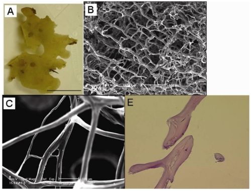
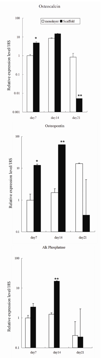
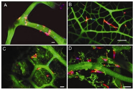
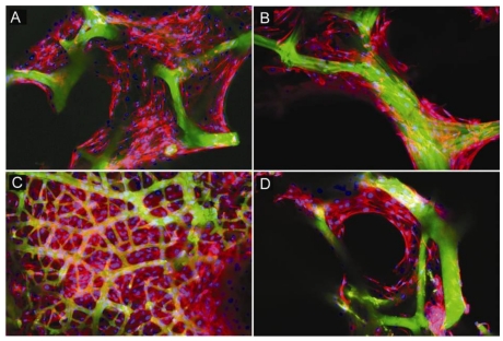
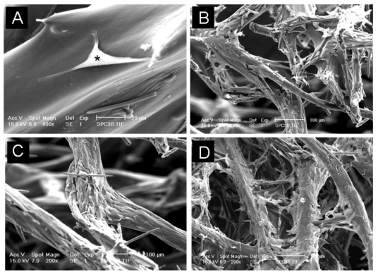
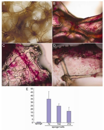
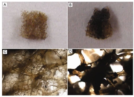
The selection of a suitable scaffold matrix is critical for cell-based bone tissue engineering. This study aimed to identify and characterize natural marine sponges as potential bioscaffolds for osteogenesis. Callyspongiidae marine sponge samples were collected from the Fremantle coast of Western Australia. The sponge structure was assessed using scanning electron microscopy (SEM) and Hematoxylin and eosin. Mouse primary osteoblasts were seeded onto the sponge scaffold and immunostained with F-actin to assess cell attachment and aggregation. Alkaline phosphatase expression, von Kossa staining and real-time PCR were performed to examine the osteogenic potential of sponge samples. SEM revealed that the sponge skeleton possessed a collagenous fibrous network consisting of interconnecting channels and a porous structure that support cellular adhesion, aggregation and growth. The average pore size of the sponge skeleton was measured 100 to 300 μm in diameter. F-actin staining demonstrated that osteoblasts were able to anchor onto the surface of collagen fibres. Alkaline phosphatase expression, a marker of early osteoblast differentiation, was evident at 7 days although expression decreased steadily with long term culture. Using von Kossa staining, mineralisation nodules were evident after 21 days. Gene expression of osteoblast markers, osteocalcin and osteopontin, was also observed at 7, 14 and 21 days of culture. Together, these results suggest that the natural marine sponge is promising as a new scaffold for use in bone tissue engineering.
选择合适的支架基质对于基于细胞的骨组织工程至关重要。本研究旨在鉴定和表征天然海洋海绵作为成骨的潜在生物支架。从西澳大利亚弗里曼特尔海岸采集了 Callispongiidae 海洋海绵样本。使用扫描电子显微镜 (SEM) 和苏木精和曙红评估海绵结构。将小鼠原代成骨细胞接种到海绵支架上,并通过 F-actin 免疫染色评估细胞附着和聚集。进行碱性磷酸酶表达、茜素红染色和实时 PCR 以检查海绵样本的成骨潜力。SEM 显示,海绵骨架具有胶原纤维状网络,由相互连接的通道和支持细胞粘附、聚集和生长的多孔结构组成。海绵骨架的平均孔径为 100 至 300μm。F-actin 染色表明成骨细胞能够附着在胶原纤维表面。碱性磷酸酶表达是成骨细胞早期分化的标志物,尽管在长期培养中表达逐渐下降,但在第 7 天就可以检测到。茜素红染色显示,在第 21 天可以看到矿化结节。在培养的第 7、14 和 21 天还观察到成骨细胞标志物骨钙素和骨桥蛋白的基因表达。这些结果表明,天然海洋海绵有望成为骨组织工程中使用的新型支架。