Department of Surgical Oncology, Osaka City University Graduate School of Medicine, 1-4-3 Asahi-machi, Abeno-ku, Osaka 545-8585, Japan.
Br J Cancer. 2011 Sep 27;105(7):996-1001. doi: 10.1038/bjc.2011.330. Epub 2011 Aug 23.
Myofibroblasts in the cancer microenvironment have recently been implicated in tumour growth and metastasis of gastric cancer. However, the mechanisms responsible for the regulation of myofibroblasts in cancer-associated fibroblasts (CAFs) remain unclear. This study was performed to clarify the mechanisms for regulation of myofibroblasts in gastric cancer microenvironment.
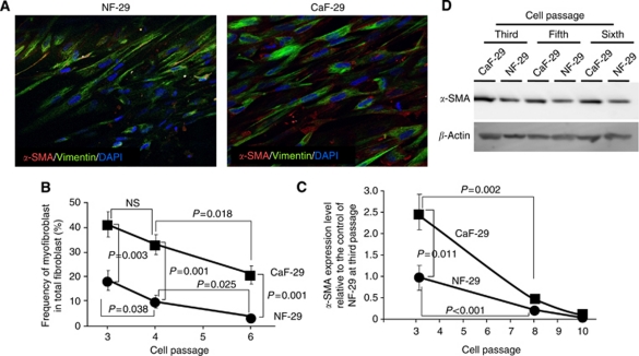
Two CAFs (CaF-29 and CaF-33) from the tumoural gastric wall and a normal fibroblast (NF-29) from the nontumoural gastric wall, 4 human gastric cancer cell lines from scirrhous gastric cancer (OCUM-2MD3 and OCUM-12), and non-scirrhous gastric cancer (MKN-45 and MKN-74) were used. Immunofluorescence microscopy by triple-immunofluorescence labelling (α-SMA, vimentin, and DAPI) was performed to determine the presence of α-SMA-positive myofibroblasts. Real-time RT-PCR was performed to examine α-SMA mRNA expression.
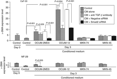
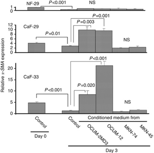
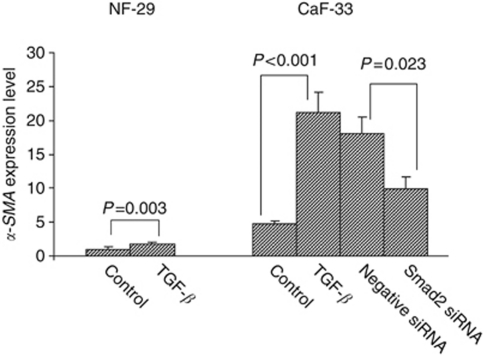
Immunofluorescence microscopy showed that the frequency of myofibroblasts in CaF-29 was greater than that in NF-29. The number of myofibroblasts in gastric fibroblasts gradually decreased with serial passages. Transforming growth factor-β (TGF-β) significantly increased the α-SMA expression level of CAFs. Conditioned medium from OCUM-2MD3 or OCUM-12 cells upregulated the α-SMA expression level of CAFs, but that from MKN-45 or MKN-74 cells did not. The α-SMA upregulation effect of conditioned medium from OCUM-2MD3 or OCUM-12 cells was significantly decreased by an anti-TGF-β antibody or Smad2 siRNA.
Transforming growth factor-β from scirrhous gastric carcinoma cells upregulates the number of myofibroblasts in CAFs.
最近有研究表明,肿瘤微环境中的肌成纤维细胞与胃癌的生长和转移有关。然而,癌相关成纤维细胞(CAFs)中肌成纤维细胞的调节机制尚不清楚。本研究旨在阐明胃癌微环境中肌成纤维细胞调节的机制。
使用来自肿瘤胃壁的两种 CAFs(CaF-29 和 CaF-33)和来自非肿瘤胃壁的正常成纤维细胞(NF-29),以及来自弥漫浸润型胃癌的 4 个人胃癌细胞系(OCUM-2MD3 和 OCUM-12)和非弥漫浸润型胃癌(MKN-45 和 MKN-74)。通过三免疫荧光标记(α-SMA、波形蛋白和 DAPI)进行免疫荧光显微镜检查,以确定α-SMA 阳性肌成纤维细胞的存在。实时 RT-PCR 用于检查α-SMA mRNA 的表达。
免疫荧光显微镜显示,CaF-29 中的肌成纤维细胞频率高于 NF-29。随着传代次数的增加,胃成纤维细胞中的肌成纤维细胞数量逐渐减少。转化生长因子-β(TGF-β)显著增加 CAFs 的α-SMA 表达水平。OCUM-2MD3 或 OCUM-12 细胞的条件培养基上调 CAFs 的α-SMA 表达水平,但 MKN-45 或 MKN-74 细胞的条件培养基没有。OCUM-2MD3 或 OCUM-12 细胞的条件培养基上调α-SMA 的作用,通过抗 TGF-β 抗体或 Smad2 siRNA 显著降低。
来自弥漫浸润型胃癌细胞的转化生长因子-β上调 CAFs 中肌成纤维细胞的数量。