Saghizadeh Mehrnoosh, Soleymani Siavash, Harounian Angel, Bhakta Bhavik, Troyanovsky Sergey M, Brunken William J, Pellegrini Graziella, Ljubimov Alexander V
Ophthalmology Research Laboratories, Cedars-Sinai Medical Center, Los Angeles, CA, USA.
Mol Vis. 2011;17:2177-90. Epub 2011 Aug 12.
We have previously identified specific epithelial proteins with altered expression in human diabetic central corneas. Decreased hepatocyte growth factor receptor (c-met) and increased proteinases were functionally implicated in the changes of these proteins in diabetes. The present study examined whether limbal stem cell marker patterns were altered in diabetic corneas and whether c-met gene overexpression could normalize these patterns.
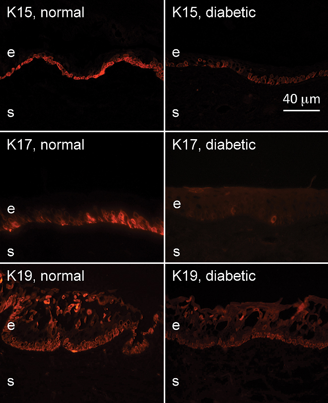
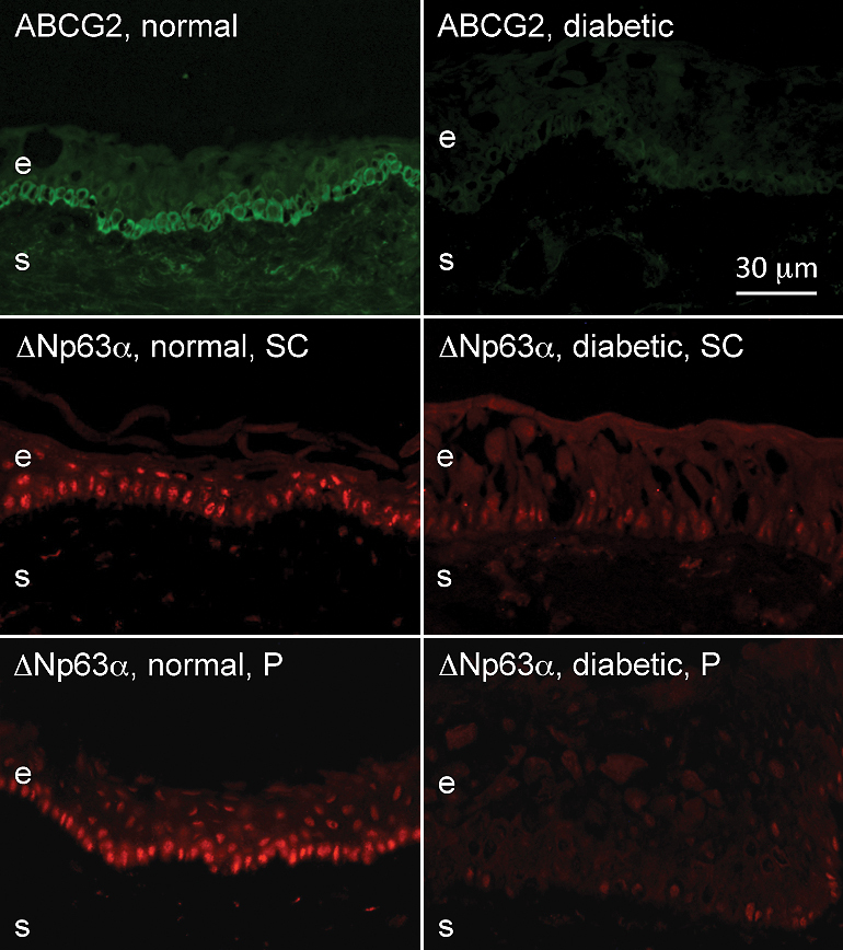
Cryostat sections of 28 ex vivo and 26 organ-cultured autopsy human normal and diabetic corneas were examined by immunohistochemistry using antibodies to putative limbal stem cell markers including ATP-binding cassette sub-family G member 2 (ABCG2), N-cadherin, ΔNp63α, tenascin-C, laminin γ3 chain, keratins (K) K15, K17, K19, β(1) integrin, vimentin, frizzled 7, and fibronectin. Organ-cultured diabetic corneas were studied upon transduction with adenovirus harboring c-met gene.
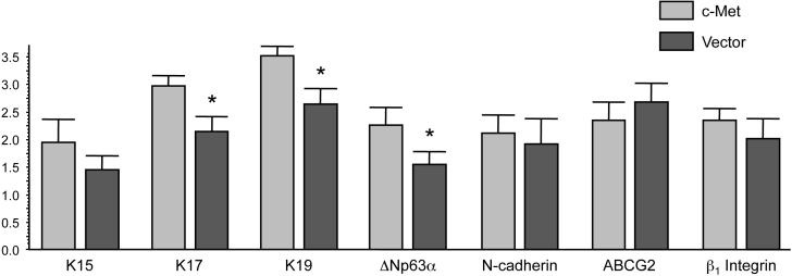
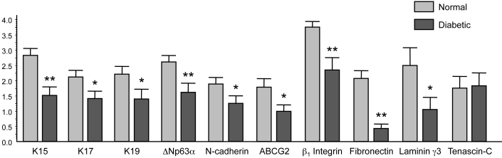
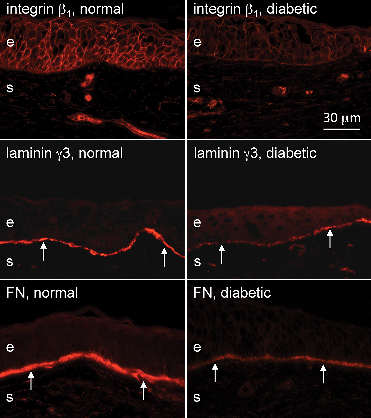
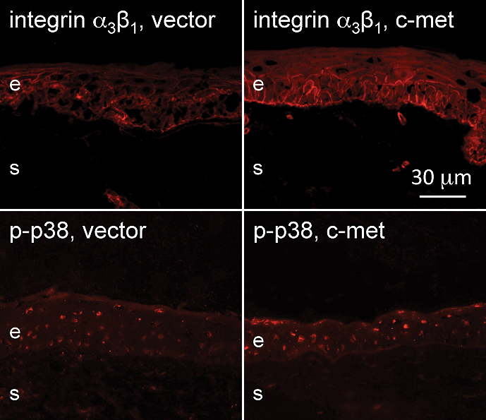
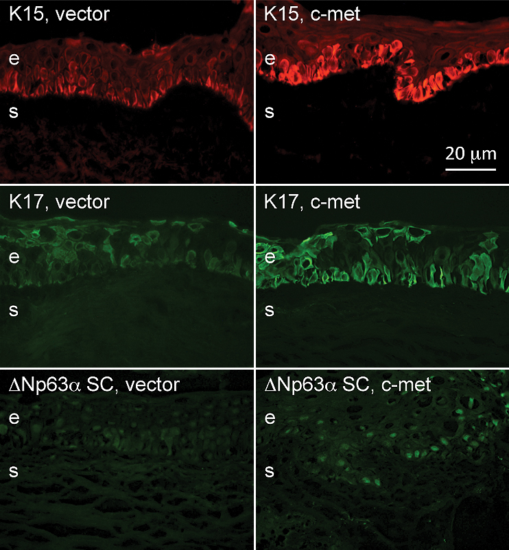
Immunostaining for ABCG2, N-cadherin, ΔNp63α, K15, K17, K19, and β(1) integrin, was significantly decreased in the stem cell-harboring diabetic limbal basal epithelium either by intensity or the number of positive cells. Basement membrane components, laminin γ3 chain, and fibronectin (but not tenascin-C) also showed a significant reduction in the ex vivo diabetic limbus. c-Met gene transduction, which normalizes diabetic marker expression and epithelial wound healing, was accompanied by increased limbal epithelial staining for K17, K19, ΔNp63α, and a diabetic marker α(3)β(1) integrin, compared to vector-transduced corneas.
The data suggest that limbal stem cell compartment is altered in long-term diabetes. Gene therapy, such as with c-met overexpression, could be able to restore normal function to diabetic corneal epithelial stem cells.
我们之前已鉴定出在人类糖尿病性中央角膜中表达发生改变的特定上皮蛋白。肝细胞生长因子受体(c-met)减少和蛋白酶增加在糖尿病中这些蛋白的变化中具有功能相关性。本研究检测糖尿病角膜中角膜缘干细胞标志物模式是否改变,以及c-met基因过表达是否能使这些模式正常化。
使用针对假定的角膜缘干细胞标志物的抗体,包括ATP结合盒亚家族G成员2(ABCG2)、N-钙黏蛋白、ΔNp63α、腱生蛋白-C、层粘连蛋白γ3链、角蛋白(K)K15、K17、K19、β(1)整合素、波形蛋白、卷曲蛋白7和纤连蛋白,通过免疫组织化学对28个离体和26个器官培养的尸检人类正常和糖尿病角膜的冰冻切片进行检测。对携带c-met基因的腺病毒转导后的器官培养糖尿病角膜进行研究。
在含有干细胞的糖尿病角膜缘基底上皮中,ABCG2、N-钙黏蛋白、ΔNp63α、K15、K17、K19和β(1)整合素的免疫染色在强度或阳性细胞数量上均显著降低。基底膜成分、层粘连蛋白γ3链和纤连蛋白(但不包括腱生蛋白-C)在离体糖尿病角膜缘中也显著减少。与载体转导的角膜相比,c-Met基因转导使糖尿病标志物表达和上皮伤口愈合正常化,同时角膜缘上皮中K17、K19、ΔNp63α和糖尿病标志物α(3)β(1)整合素的染色增加。
数据表明长期糖尿病会改变角膜缘干细胞区室。基因治疗,如c-met过表达,可能能够恢复糖尿病角膜上皮干细胞的正常功能。