Department of Cardiovascular Medicine and Oxford Acute Vascular Imaging Centre, University of Oxford, John Radcliffe Hospital, Oxford OX3 9DU, United Kingdom.
Atherosclerosis. 2011 Dec;219(2):579-87. doi: 10.1016/j.atherosclerosis.2011.07.127. Epub 2011 Aug 5.
Optical coherence tomography (OCT) is a high resolution imaging technique used to assess superficial atherosclerotic plaque morphology. Utility of OCT may be enhanced by contrast agents targeting molecular mediators of inflammation.
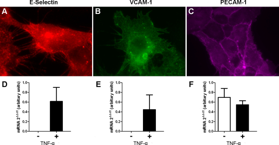
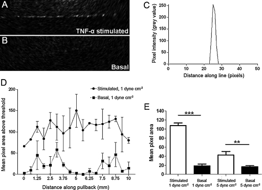
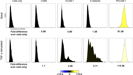
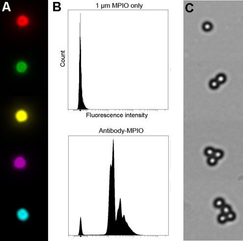
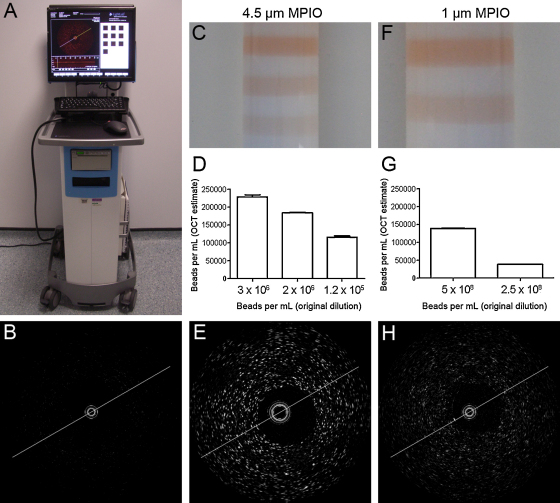
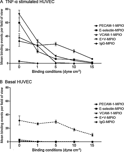
Microparticles of iron oxide (MPIO; 1 and 4.5 μm diameter) in suspension were visualized and accurately quantified using a clinical optical coherence tomography system. Bound to PECAM-1 on a plane of cultured endothelial cells under static conditions, 1 μm MPIO were also readily detected by OCT. To design a molecular contrast probe that would bind activated endothelium under conditions of shear stress, we quantified the expression (basal vs. TNF-activated; molecules μm(-2)) of VCAM-1 (not detected vs. 16 ± 1); PECAM-1 (132 ± 6 vs. 198 ± 10) and E-selectin (not detected vs. 46 ± 0.6) using quantitative flow cytometry. We then compared the retention of antibody-conjugated MPIO targeting each of these molecules plus a combined VCAM-1 and E-selectin (E+V) probe across a range of physiologically relevant shear stresses. E+V MPIO were consistently retained with highest efficiency (P < 0.001) and at a density that provided conspicuous contrast effects on OCT pullback.
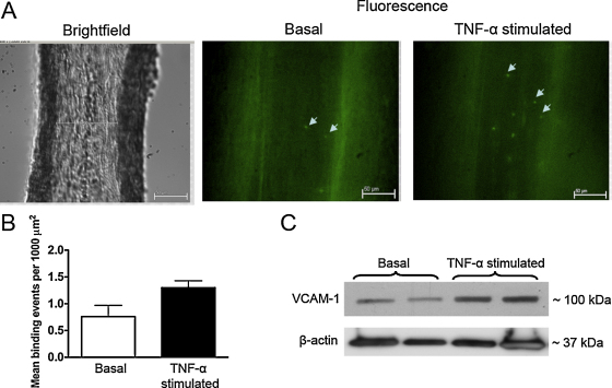
Microparticles of iron oxide were detectable using a clinical OCT system. Assessment of binding under flow conditions recommended an approach that targeted both E-selectin and VCAM-1. Bound to HUVEC under conditions of flow, targeted 1 μm E+V MPIO were readily identified on OCT pullback. Molecular imaging with OCT may be feasible in vivo using antibody targeted MPIO.
光学相干断层扫描(OCT)是一种用于评估表面动脉粥样硬化斑块形态的高分辨率成像技术。通过靶向炎症分子介质的造影剂,可增强 OCT 的效用。
悬浮的氧化铁微颗粒(MPIO;1 和 4.5μm 直径)可使用临床光学相干断层扫描系统进行可视化和准确量化。在培养的内皮细胞平面上在静态条件下与 PECAM-1 结合,1μm MPIO 也可通过 OCT 轻松检测到。为了设计一种在切应力条件下结合活化内皮细胞的分子对比探针,我们定量了 VCAM-1(未检测到 vs. 16±1)、PECAM-1(132±6 vs. 198±10)和 E-选择素(未检测到 vs. 46±0.6)在基础与 TNF 激活条件下的表达(分子/μm²)。然后,我们比较了针对这些分子中的每一个以及 VCAM-1 和 E-选择素(E+V)探针的抗体偶联 MPIO 的保留情况,范围涵盖了一系列生理相关的切应力。E+V MPIO 以最高效率(P<0.001)和提供 OCT 回缩时明显对比效果的密度一致地保留下来。
临床 OCT 系统可检测氧化铁微颗粒。在流动条件下的结合评估建议采用靶向 E-选择素和 VCAM-1 的方法。在流动条件下与 HUVEC 结合,靶向 1μm E+V MPIO 可在 OCT 回缩时轻松识别。使用抗体靶向 MPIO 可能可在体内进行 OCT 分子成像。