Scientific & Medical Affairs, Sigma Tau SpA, Pomezia, 00040, Roma, Italy.
Curr Neuropharmacol. 2011 Mar;9(1):195-9. doi: 10.2174/157015911795017182.
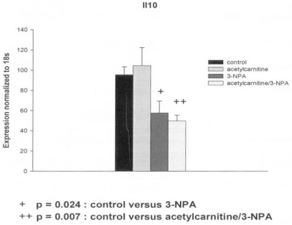
The neurotoxicity induced by the mitochondrial inhibitor 3-nitropropionic acid (3-NPA) is associated with a decrease of ATP synthesis and an increase of free radical production which can lead to apoptosis or necrosis. We have used the PC12, neuron-like rat pheochromocytoma cell line, to study further the mechanism of 3-NPA-evoked neurotoxicity and the effects of acetyl-L-carnitine (ALC) which has neuroprotective actions against various types of mitochondrial inhibitors.Cultured PC 12 cells were exposed to a low dose of 3-NPA 50 (microM) in the presence or absence of 5 mM ALC. The dose of 3-NPA was sub toxic and no changes in pro-apoptotic Bax or anti-apoptotic Bcl-2 gene expression were observed. We followed specific genetic markers to look for changes evoked by 3-NPA toxicity and also changes associated with neuroprotection exerted by the ALC treatment, using RT-PCR arrays (delta-delta method). 3-NPA exposure evoked a decrease in expression of the Tp53 gene. This down regulation was prevented by pretreatment of the cells with ALC. The Tp53 gene responds to cellular stresses and the effects seen here are possibly associated with the 3-NPA evoked changes in mitochondrial metabolism. Other genes associated with stress and apoptosis, Parp-1, Bcl-2, and Bax were not affected by 3-NPA or ALC. The decrease of inflammatory response Il-10 gene expression due to 3-NPA was further lowered by presence of ALC. Other inflammation related genes, Il1rn, Nr3c1 and Cxcr4 were not affected. Interestingly, the glutamate transporter slc17a7, carnitine-acylcarnitine translocase Slc25a20 and heat shock proteins genes, Hsp27, Hmox1 (Hsp32, HO1) as well as Hspa 1a (Hsp 70) increased only when both ALC and small dose of 3-NPA were present. The alterations in gene expression detected in this study suggest role of several intracellular pathways in the neurotoxicity of 3-NPA and the neuroprotection against 3-NPA-induced neurotoxicity by ALC.
线粒体抑制剂 3-硝基丙酸(3-NPA)引起的神经毒性与 ATP 合成减少和自由基产生增加有关,这可能导致细胞凋亡或坏死。我们使用 PC12 细胞(大鼠嗜铬细胞瘤神经元样细胞系)进一步研究 3-NPA 诱导的神经毒性的机制,以及乙酰左旋肉碱(ALC)的作用。ALC 对各种类型的线粒体抑制剂具有神经保护作用。
将培养的 PC12 细胞暴露于低剂量的 3-NPA(50μM),存在或不存在 5mM 的 ALC。3-NPA 的剂量是亚毒性的,促凋亡 Bax 或抗凋亡 Bcl-2 基因表达没有变化。我们使用 RT-PCR 阵列(delta-delta 方法),跟踪特定的遗传标记,寻找 3-NPA 毒性引起的变化,以及与 ALC 处理的神经保护作用相关的变化。3-NPA 暴露引起 Tp53 基因表达下调。这种下调可被细胞的 ALC 预处理所阻止。Tp53 基因对细胞应激作出反应,这里观察到的效应可能与 3-NPA 引起的线粒体代谢变化有关。其他与应激和细胞凋亡相关的基因,Parp-1、Bcl-2 和 Bax 不受 3-NPA 或 ALC 的影响。由于 3-NPA 引起的抗炎反应 Il-10 基因表达的降低,由于 ALC 的存在而进一步降低。其他与炎症相关的基因,Il1rn、Nr3c1 和 Cxcr4 不受影响。有趣的是,只有当同时存在 ALC 和小剂量的 3-NPA 时,谷氨酸转运体 slc17a7、肉碱酰基辅酶 A 转移酶 Slc25a20 和热休克蛋白基因 Hsp27、Hmox1(Hsp32,HO1)以及 Hspa1a(Hsp70)才会增加。本研究中检测到的基因表达变化表明,几种细胞内途径在 3-NPA 的神经毒性和 ALC 对抗 3-NPA 诱导的神经毒性的神经保护作用中发挥作用。