Sankhla Suresh, Khan G M
Department of Neurosurgery, Dr. Balabhai Nanavati Hospital, Mumbai, India.
J Pediatr Neurosci. 2009 Jan;4(1):2-9. doi: 10.4103/1817-1745.49098.
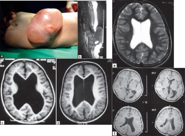
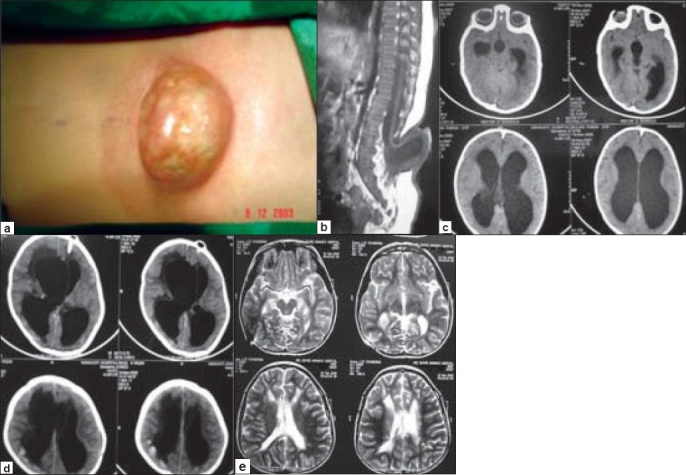
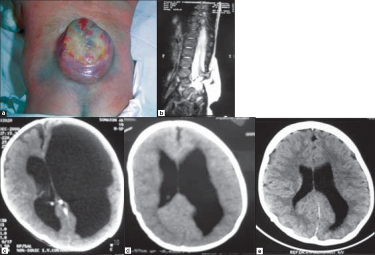
The incidence of hydrocephalus requiring shunts in children with myelomeningocele (MMC) is reported to be very high. Shunt-related complications are a significant cause of morbidity and mortality in this population. In order to minimize shunt placements, we used very rigid clinical selection criteria and followed them in all patients who had myelomeningocele and enlarged ventricles. The follow-up outcome of this retrospective study is reported.
From 2000 to 2007, 23 patients with myelomeningocele and variable degree of hydrocephalus were treated at our institute with primary surgical closure of their myelomeningoceles without a CSF diversion procedure. Patients with severe hydrocephalus who required immediate shunt insertion, and those with no significant associated hydrocephalus were not included in this study. Data regarding the surgical results and complications, postoperative management, and the outcome at follow-up were obtained from their hospital records.
Initially increased size of the ventricular system was found to have decreased or stabilized in 17 (81%) patients postoperatively. However, ventriculomegaly continued to progress further in 4 (19%) out of 21 patients. Of 11 patients who presented with enlarged head, eight (73%) patients showed reduction or stabilization in their head circumference. Three (27%) children continued to have progressive head enlargement in the postoperative period and required shunt placement. Signs of raised intracranial pressure observed in six patients on admission, improved in two (33%) and persisted or worsened in four (67%) patients who eventually improved after the insertion of a shunt. Eight (35%) patients experienced wound-related complications following closure of the MMC, including CSF leak in four, wound infection in three, wound breakdown in three, and pseudomeningocele in two patients. Shunt placement was required in the postoperative period in 13 (56.5%) patients to treat raised intracranial pressure in 11 and CSF leak from the wound in two patients.
Our experience suggests that the placement of shunts can be reduced by adopting a policy with strict clinical and radiographic criteria. Shunt insertion should be reserved for only those patients who have severe hydrocephalus with clinical features of elevated intracranial pressure. Mild to moderate ventricular dilatation, persistent ventriculomagaly, and some increase in ventricular size after myelomeningocele repair can be treated successfully without a shunt.
据报道,脊髓脊膜膨出(MMC)患儿中需要分流术治疗脑积水的发生率非常高。分流相关并发症是该人群发病和死亡的重要原因。为了尽量减少分流术的实施,我们采用了非常严格的临床选择标准,并在所有患有脊髓脊膜膨出和脑室扩大的患者中遵循这些标准。本文报告了这项回顾性研究的随访结果。
2000年至2007年期间,我院对23例患有脊髓脊膜膨出且脑积水程度不一的患者进行了治疗,初次手术闭合其脊髓脊膜膨出,未进行脑脊液分流手术。需要立即插入分流管的严重脑积水患者以及无明显相关脑积水的患者未纳入本研究。从患者的医院记录中获取有关手术结果和并发症、术后管理以及随访结果的数据。
术后发现17例(81%)患者最初增大的脑室系统缩小或稳定。然而,21例患者中有4例(19%)脑室扩大继续进展。11例头围增大的患者中,8例(73%)患者的头围缩小或稳定。3例(27%)儿童术后头围继续进行性增大,需要进行分流术。6例入院时出现颅内压升高体征的患者中,2例(33%)有所改善,4例(67%)患者的体征持续或恶化,最终在插入分流管后有所改善。8例(35%)患者在脊髓脊膜膨出闭合后出现伤口相关并发症,包括4例脑脊液漏、3例伤口感染、3例伤口裂开和2例假性脑脊膜膨出。术后13例(56.5%)患者需要进行分流术,其中11例用于治疗颅内压升高,2例用于治疗伤口脑脊液漏。
我们的经验表明,通过采用严格的临床和影像学标准的策略,可以减少分流术的实施。分流术应仅保留给那些患有严重脑积水且具有颅内压升高临床特征的患者。轻度至中度脑室扩张、持续性脑室扩大以及脊髓脊膜膨出修复后脑室大小的一些增加可以在不进行分流术的情况下成功治疗。