INSERM, U892, Université de Nantes, Nantes, France.
PLoS Pathog. 2011 Aug;7(8):e1002188. doi: 10.1371/journal.ppat.1002188. Epub 2011 Aug 25.
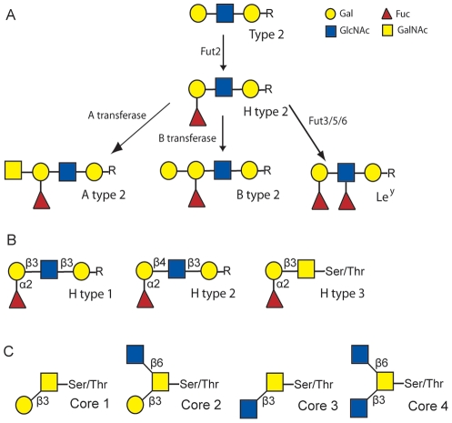
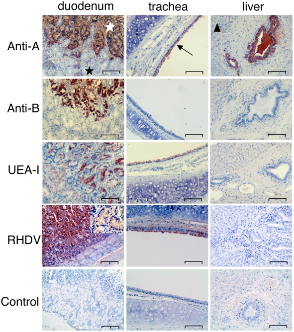
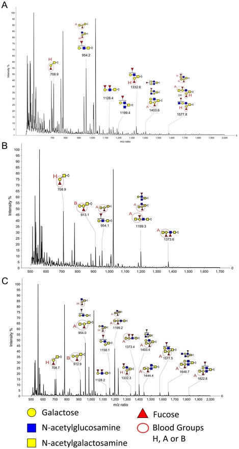
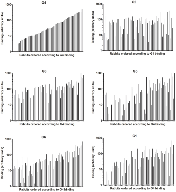
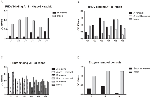
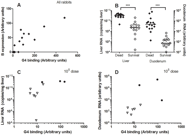
Rabbit Hemorrhagic disease virus (RHDV), a calicivirus of the Lagovirus genus, and responsible for rabbit hemorrhagic disease (RHD), kills rabbits between 48 to 72 hours post infection with mortality rates as high as 50-90%. Caliciviruses, including noroviruses and RHDV, have been shown to bind histo-blood group antigens (HBGA) and human non-secretor individuals lacking ABH antigens in epithelia have been found to be resistant to norovirus infection. RHDV virus-like particles have previously been shown to bind the H type 2 and A antigens. In this study we present a comprehensive assessment of the strain-specific binding patterns of different RHDV isolates to HBGAs. We characterized the HBGA expression in the duodenum of wild and domestic rabbits by mass spectrometry and relative quantification of A, B and H type 2 expression. A detailed binding analysis of a range of RHDV strains, to synthetic sugars and human red blood cells, as well as to rabbit duodenum, a likely gastrointestinal site for viral entrance was performed. Enzymatic cleavage of HBGA epitopes confirmed binding specificity. Binding was observed to blood group B, A and H type 2 epitopes in a strain-dependent manner with slight differences in specificity for A, B or H epitopes allowing RHDV strains to preferentially recognize different subgroups of animals. Strains related to the earliest described RHDV outbreak were not able to bind A, whereas all other genotypes have acquired A binding. In an experimental infection study, rabbits lacking the correct HBGA ligands were resistant to lethal RHDV infection at low challenge doses. Similarly, survivors of outbreaks in wild populations showed increased frequency of weak binding phenotypes, indicating selection for host resistance depending on the strain circulating in the population. HBGAs thus act as attachment factors facilitating infection, while their polymorphism of expression could contribute to generate genetic resistance to RHDV at the population level.
兔出血症病毒(RHDV)是杯状病毒科兔病毒属的成员,可引起兔出血症(RHD),感染后 48 至 72 小时内兔子死亡率高达 50-90%。杯状病毒,包括诺如病毒和 RHDV,已被证明可以结合组织血型抗原(HBGA),而上皮组织中缺乏 ABH 抗原的人类非分泌者已被发现对诺如病毒感染具有抵抗力。RHDV 病毒样颗粒以前被证明可以结合 H 型 2 和 A 抗原。在这项研究中,我们全面评估了不同 RHDV 分离株对 HBGA 的株特异性结合模式。我们通过质谱法和 A、B 和 H 型 2 表达的相对定量来表征野生和家兔十二指肠中的 HBGA 表达。对一系列 RHDV 株与合成糖和人类红细胞以及兔十二指肠的详细结合分析表明,病毒进入的胃肠道部位可能是十二指肠。HBGA 表位的酶切确认了结合的特异性。观察到以菌株依赖的方式与血型 B、A 和 H 型 2 表位结合,对 A、B 或 H 表位的特异性略有差异,使 RHDV 株能够优先识别不同的动物亚群。与最早描述的 RHDV 暴发相关的菌株不能结合 A,而所有其他基因型都获得了 A 结合。在实验感染研究中,缺乏正确 HBGA 配体的兔子在低剂量挑战时对致死性 RHDV 感染具有抗性。同样,在野生种群暴发的幸存者中,弱结合表型的出现频率增加,表明随着流行株的变化,宿主抗性选择。因此,HBGA 作为附着因子促进感染,而其表达的多态性可能有助于在群体水平上产生对 RHDV 的遗传抗性。