Baroncini Liz Andréa V, Filho Pazin Antonio, Murta Luiz Otávio, Nakao Lia S, Ramos Simone G, Précoma Dalton B
Department of Health and Scienses - Pontificia Universidade Católica do Paraná, Rua Imaculada Conceição 1155, Curitiba, Paraná, CEP: 80215901, Brazil.
Cardiovasc Ultrasound. 2011 Sep 18;9:24. doi: 10.1186/1476-7120-9-24.
Matrix metalloproteinase-9 (MMP-9) and tissue inhibitor of MMP (TIMP) promote derangement of the extracellular matrix, which is ultimately reflected in plaque images seen on ultrasound. Videodensitometry can identify structural disturbances in plaques.
To establish the correlations between values determined using videodensitometry in B-mode ultrasound images of advanced carotid plaques and the total expression of MMP-9 and TIMP-1 in these removed plaques.
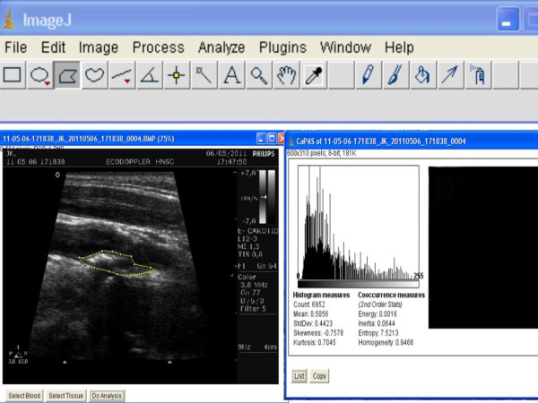
Thirty patients underwent ultrasonic tissue characterization of carotid plaques before surgery, using mean gray level (MGL), energy, entropy and homogeneity. Each patient was assigned preoperatively to one of 2 groups: group I, symptomatic patients (n = 16; 12 males; mean age 66.7 ± 6.8 years), and group II, asymptomatic patients (n = 14; 8 males; mean age 67.6 ± 6.81 years). Tissue specimens were analyzed for MMP-9 and TIMP-1 expression. Nine carotid arteries were used as normal tissue controls.
MMP-9 expression levels were elevated in group II and in normal tissues compared to group I (p < 0.001). TIMP-1 levels were higher in group II than in group I, and significantly higher in normal tissues than in group I (p = 0.039). The MGL was higher in group II compared to group I (p = 0.038). Energy had greater values in group II compared to group I (p = 0.02). There were no differences between patient groups in homogeneity and entropy. Energy positively correlated with MMP-9 and TIMP-1 expression (p = 0.012 and p = 0.031 respectively). Homogeneity positively correlated with MMP-9 and TIMP-1 expression (p = 0.034 and p = 0.047 respectively). There were no correlations between protein expression and MGL or entropy.
Videodensitometric computer analysis of ultrasound scanning images can be used to identify stable carotid plaques, which have higher total expression levels of MMP-9 and TIMP-1 than unstable plaques.
基质金属蛋白酶-9(MMP-9)和MMP组织抑制剂(TIMP)会促使细胞外基质紊乱,这最终会反映在超声检查中看到的斑块图像上。视频密度测定法可以识别斑块中的结构紊乱。
确定在晚期颈动脉斑块的B型超声图像中使用视频密度测定法所测定的值与这些切除斑块中MMP-9和TIMP-1的总表达之间的相关性。
30例患者在手术前接受了颈动脉斑块的超声组织特征分析,采用平均灰度值(MGL)、能量、熵和均匀性指标。每位患者在术前被分为2组之一:I组,有症状患者(n = 16;男性12例;平均年龄66.7±6.8岁),II组,无症状患者(n = !4;男性8例;平均年龄67.6±6.81岁)。对组织标本进行MMP-9和TIMP-1表达分析。9条颈动脉用作正常组织对照。
与I组相比,II组和正常组织中MMP-9表达水平升高(p < 0.001)。II组中TIMP-1水平高于I组,正常组织中TIMP-1水平显著高于I组(p = 0.039)。II组的MGL高于I组(p = 0.038)。II组的能量值高于I组(p = 0.02)。患者组之间在均匀性和熵方面没有差异。能量与MMP-9和TIMP-1表达呈正相关(分别为p = 0.012和p = 0.031)。均匀性与MMP-9和TIMP-1表达呈正相关(分别为p = 0.034和p = 0.047)。蛋白质表达与MGL或熵之间没有相关性。
超声扫描图像的视频密度测定计算机分析可用于识别稳定的颈动脉斑块,其MMP-9和TIMP-1的总表达水平高于不稳定斑块。