Siwicka Karolina A, Kitoh Hiroshi, Kawasumi Motoaki, Ishiguro Naoki
Nagoya University Graduate School of Medicine, Department of Orthopaedic Surgery, Nagoya, Japan.
Nagoya J Med Sci. 2011 Aug;73(3-4):117-27.
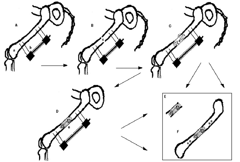
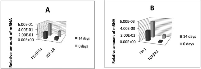
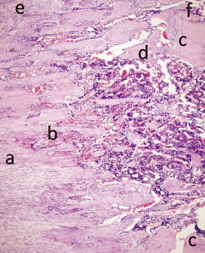
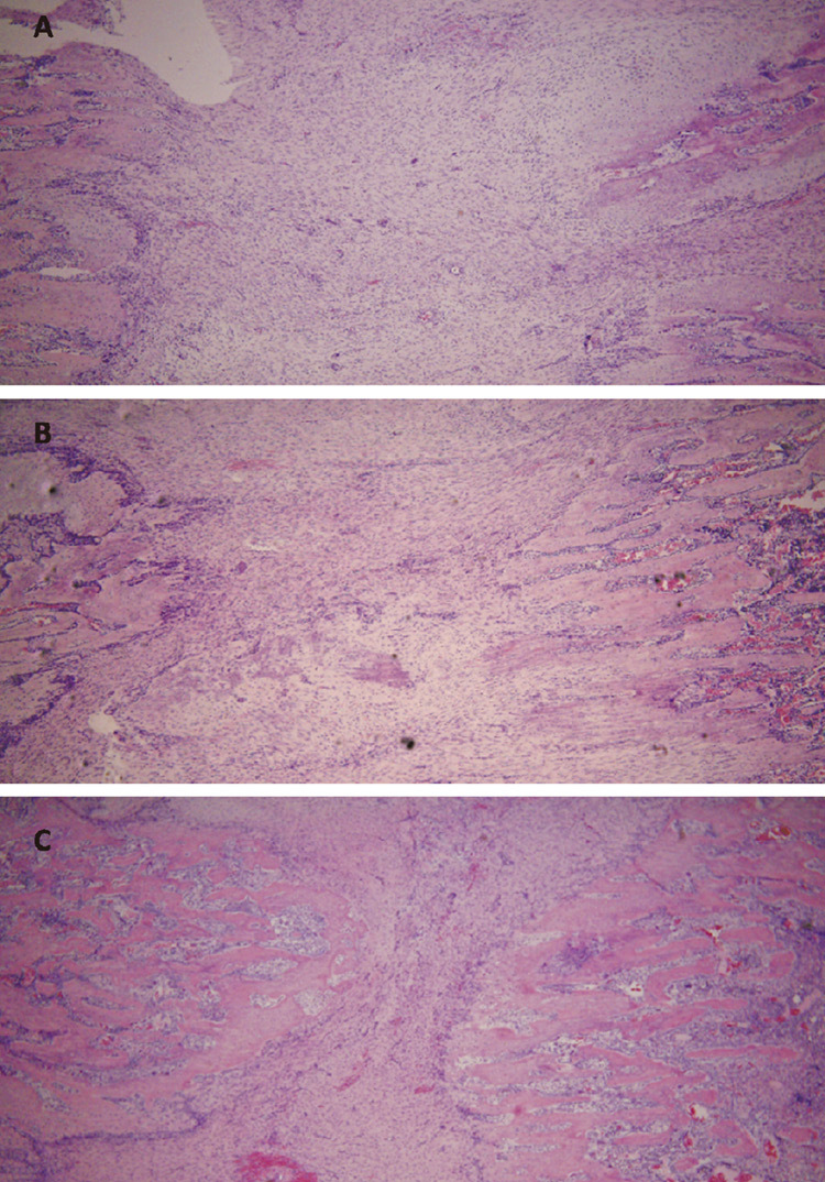
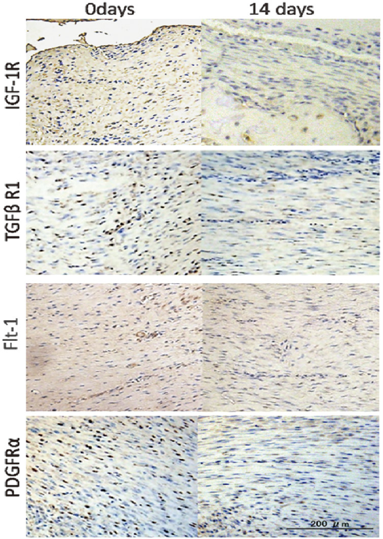
Management of bone deficits by distraction osteogenesis is an appreciated but lengthy procedure. To accelerate the consolidation of newly formed distraction callus, an administration of growth factors into the distraction gap has been suggested. Changes in expression of growth factors receptors in the distracted callus during consolidation were studied in order to improve our understanding of the underlying molecular mechanisms and to provide a scientific basis for clinical application of growth factors. In a model of rat bone lengthening the expression of receptors for: vascular endothelial growth factor, transforming growth factor beta1, insulin like growth factor and platelet derived growth factor were evaluated semiquantitatively with immunohistochemistry and quantitatively with real time PCR in various callus zones at zero, one and two weeks of consolidation. Overall growth factors receptors' expression was highest at the beginning of consolidation. It was strongest in the trabecular bone and weakest in the fibrous zone. Transforming growth factor beta receptor 1 was most abundant and vascular endothelial growth factor receptor 1, although scarce, showed the most consistent expression. In contrast to the osteogenic zones, the fibrous zone demonstrated a dramatic loss of the growth factors receptors over time. High growth factors receptors expression shortly after termination of the distraction may warrant the maximal callus' response to injected growth factors. Rapid decline of growth factors receptors in the fibrous zone may imply its decreasing sensitivity to growth factors and, as a consequence, a declining osteogenic potential.
通过牵张成骨术治疗骨缺损是一种公认但耗时的方法。为了加速新形成的牵张骨痂的巩固,有人建议向牵张间隙内注入生长因子。研究了骨痂在巩固过程中牵张部位生长因子受体表达的变化,以增进我们对潜在分子机制的理解,并为生长因子的临床应用提供科学依据。在大鼠骨延长模型中,通过免疫组织化学半定量和实时聚合酶链反应定量评估了在巩固的第零周、第一周和第二周时,不同骨痂区域中血管内皮生长因子、转化生长因子β1、胰岛素样生长因子和血小板衍生生长因子受体的表达。总体而言,生长因子受体的表达在巩固开始时最高。在小梁骨中最强,在纤维区最弱。转化生长因子β受体1最为丰富,血管内皮生长因子受体1虽然稀少,但表达最为一致。与成骨区相比,纤维区的生长因子受体随时间急剧减少。牵张结束后不久生长因子受体的高表达可能保证了骨痂对注射生长因子的最大反应。纤维区生长因子受体的快速下降可能意味着其对生长因子的敏感性降低,因此成骨潜力下降。