Department of Biomedical Engineering, Tufts University, Medford, Massachusetts, United States of America.
PLoS One. 2011;6(9):e24765. doi: 10.1371/journal.pone.0024765. Epub 2011 Sep 9.
Multi-photon fluorescence microscopy techniques allow for non-invasive interrogation of live samples in their native environment. These methods are particularly appealing for identifying pre-cancers because they are sensitive to the early changes that occur on the microscopic scale and can provide additional information not available using conventional screening techniques.
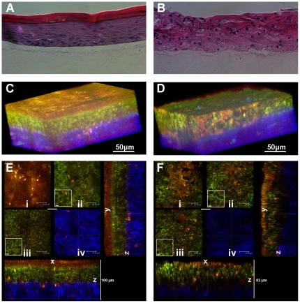
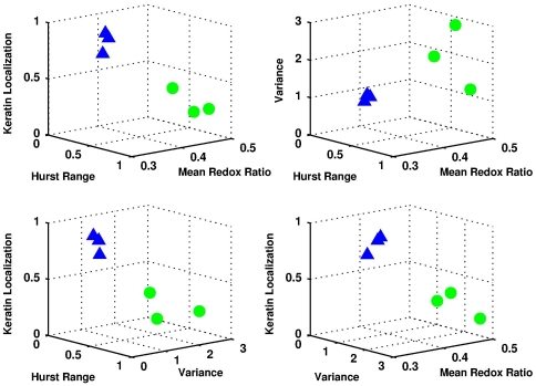
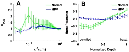
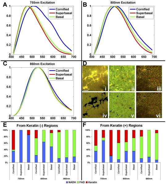
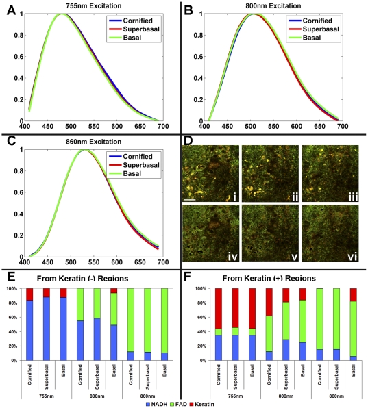
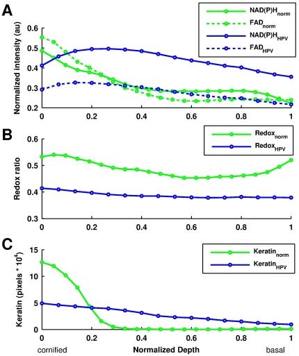
METHODOLOGY/PRINCIPAL FINDINGS: In this study, we developed novel automated approaches, which can be employed for the real-time analysis of two-photon fluorescence images, to non-invasively discriminate between normal and pre-cancerous/HPV-immortalized engineered tissues by concurrently assessing metabolic activity, morphology, organization, and keratin localization. Specifically, we found that the metabolic activity was significantly enhanced and more uniform throughout the depths of the HPV-immortalized epithelia, based on our extraction of the NADH and FAD fluorescence contributions. Furthermore, we were able to separate the keratin contribution from metabolic enzymes to improve the redox estimates and to use the keratin localization as a means to discriminate between tissue types. To assess morphology and organization, Fourier-based, power spectral density (PSD) approaches were employed. The nuclear size distribution throughout the epithelial depths was quantified by evaluating the variance of the corresponding spatial frequencies, which was found to be greater in the normal tissue compared to the HPV-immortalized tissues. The PSD was also used to calculate the Hurst parameter to identify the level of organization in the tissues, assuming a fractal model for the fluorescence intensity fluctuations within a field. We found the range of organization was greater in the normal tissue and closely related to the level of differentiation.
CONCLUSIONS/SIGNIFICANCE: A wealth of complementary morphological, biochemical and organizational tissue parameters can be extracted from high resolution images that are acquired based entirely on endogenous sources of contrast. They are promising diagnostic parameters for the non-invasive identification of early cancerous changes and could improve significantly diagnosis and treatment for numerous patients.
多光子荧光显微镜技术允许在其自然环境中对活样本进行非侵入性询问。这些方法对于识别癌前病变特别有吸引力,因为它们对微观尺度上发生的早期变化敏感,并且可以提供使用传统筛选技术无法获得的附加信息。
方法/主要发现:在这项研究中,我们开发了新的自动化方法,可以用于实时分析双光子荧光图像,通过同时评估代谢活性、形态、组织和角蛋白定位,非侵入性地区分正常和癌前病变/HPV 永生化工程组织。具体来说,我们发现基于 NADH 和 FAD 荧光贡献的提取,HPV 永生化上皮组织的代谢活性显著增强且更加均匀。此外,我们能够将角蛋白从代谢酶中分离出来,以改善氧化还原估计,并使用角蛋白定位作为区分组织类型的手段。为了评估形态和组织,我们采用了基于傅里叶的功率谱密度(PSD)方法。通过评估相应空间频率的方差,定量评估了上皮深度内的核大小分布,发现正常组织中的方差大于 HPV 永生化组织。PSD 还用于计算 Hurst 参数,以识别组织中的组织水平,假设荧光强度波动的分形模型在一个场中。我们发现正常组织的组织范围更大,并且与分化水平密切相关。
结论/意义:可以从完全基于内源性对比源获取的高分辨率图像中提取出丰富的形态、生化和组织参数。它们是用于非侵入性识别早期癌变变化的有前途的诊断参数,并可以显著改善许多患者的诊断和治疗。