Institute for Molecular Cardiovascular Research, University Hospital Aachen, Rheinisch-Westfälische Technische Hochschule Aachen, Aachen, Germany.
Cell Death Dis. 2011 Sep 29;2(9):e211. doi: 10.1038/cddis.2011.94.
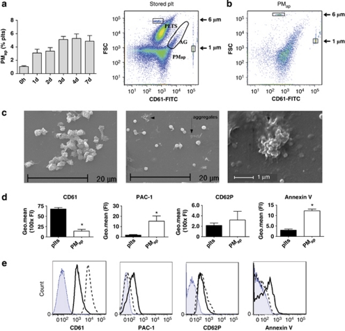
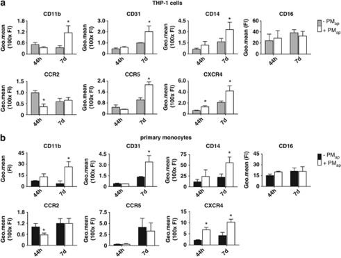
Platelets shed microparticles not only upon activation, but also upon ageing by an apoptosis-like process (apoptosis-induced platelet microparticles, PM(ap)). While the activation-induced microparticles have widely been studied, not much is known about the (patho)physiological consequences of PM(ap) formation. Flow cytometry and scanning electron microscopy demonstrated that PM(ap) display activated integrins and interact to form microparticle aggregates. PM(ap) were chemotactic for monocytic cells, bound to these cells, an furthermore stimulated cell adhesion and spreading on a fibronectin surface. After prolonged incubation, PM(ap) promoted cell differentiation, but inhibited proliferation. Monocyte membrane receptor analysis revealed increased expression levels of CD11b (integrin α(M)β(2)), CD14 and CD31 (platelet endothelial cell adhesion molecule-1), and the chemokine receptors CCR5 and CXCR4, but not of CCR2. This indicated that PM(ap) polarized the cells into resident M2 monocytes. Cells treated with PM(ap) actively consumed oxidized low-density lipoprotein (oxLDL), and released matrix metalloproteinases and hydrogen peroxide. Further confirmation for the differentiation towards resident professional phagocytes came from the finding that PM(ap) stimulated the expression of the (ox)LDL receptors, CD36 and CD68, and the production of proinflammatory and immunomodulating cytokines by monocytes. In conclusion, interaction of PM(ap) with monocytic cells has an immunomodulating potential. The apoptotic microparticles polarize the cells into a resident M2 subset, and induce differentiation to resident professional phagocytes.
血小板不仅在激活时释放微颗粒,而且在凋亡样过程(凋亡诱导的血小板微颗粒,PM(ap))中也会释放微颗粒。虽然已经广泛研究了激活诱导的微颗粒,但对 PM(ap)形成的(病理)生理后果知之甚少。流式细胞术和扫描电子显微镜显示,PM(ap)显示出激活的整合素并相互作用形成微颗粒聚集。PM(ap)对单核细胞具有趋化性,与这些细胞结合,并进一步刺激细胞在纤维连接蛋白表面上的黏附和铺展。经过长时间孵育,PM(ap)促进细胞分化,但抑制增殖。单核细胞膜受体分析显示 CD11b(整合素 α(M)β(2))、CD14 和 CD31(血小板内皮细胞黏附分子-1)以及趋化因子受体 CCR5 和 CXCR4 的表达水平增加,但 CCR2 的表达水平没有增加。这表明 PM(ap)将细胞极化成为驻留的 M2 单核细胞。用 PM(ap)处理的细胞积极消耗氧化低密度脂蛋白 (oxLDL),并释放基质金属蛋白酶和过氧化氢。PM(ap)刺激单核细胞表达 (ox)LDL 受体 CD36 和 CD68 以及产生促炎和免疫调节细胞因子的发现进一步证实了向驻留的专业吞噬细胞的分化。总之,PM(ap)与单核细胞的相互作用具有免疫调节潜力。凋亡的微颗粒将细胞极化成为驻留的 M2 亚群,并诱导分化为驻留的专业吞噬细胞。