Yamana Ippei, Kawamoto Shunji, Nagao Shuji, Yoshida Takahisa, Yamashita Yuichi
Department of Surgery, Fukuoka University School of Medicine, Fukuoka, Japan.
Case Rep Gastroenterol. 2011 May;5(2):463-70. doi: 10.1159/000331051. Epub 2011 Aug 22.
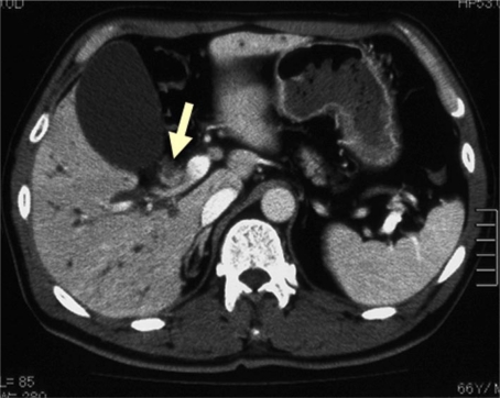
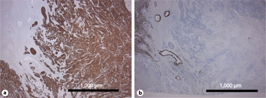
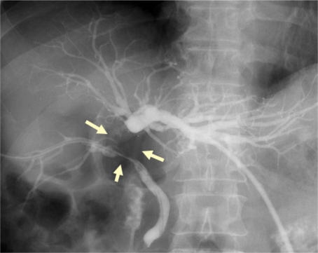
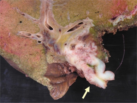
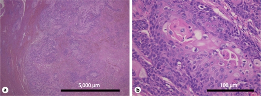
We herein report a rare case of squamous cell carcinoma of the hilar bile duct. A 66-year-old Japanese male patient was admitted to our hospital because of appetite loss and jaundice. Abdominal computed tomography revealed an enhanced mass measuring 10 × 30 mm in the hilar bile duct region. After undergoing biliary drainage, the patient underwent extended right hepatic lobectomy with regional lymph nodes dissection. The tumor had invaded the right portal vein. Therefore, we also performed resection and reconstruction of the portal vein. Histopathologically, the carcinoma cells exhibited a solid structure with differentiation to squamous cell carcinoma with keratinization and intercellular bridges. Immunohistochemical staining of the tumor cells revealed positive cytokeratin staining and negative CAM 5.2 staining. Based on these findings, a definitive diagnosis of well-differentiated squamous cell carcinoma of the hilar bile duct was made.
我们在此报告一例罕见的肝门部胆管鳞状细胞癌病例。一名66岁的日本男性患者因食欲减退和黄疸入院。腹部计算机断层扫描显示肝门部胆管区域有一个大小为10×30毫米的强化肿块。在进行胆道引流后,患者接受了扩大右肝叶切除术并进行区域淋巴结清扫。肿瘤侵犯了右门静脉。因此,我们还进行了门静脉切除和重建。组织病理学检查显示,癌细胞呈实体结构,分化为伴有角化和细胞间桥的鳞状细胞癌。肿瘤细胞的免疫组织化学染色显示细胞角蛋白染色阳性,CAM 5.2染色阴性。基于这些发现,确诊为肝门部胆管高分化鳞状细胞癌。