• 文献检索
  • 文档翻译
  • 深度研究
  • 学术资讯
  • Suppr Zotero 插件Zotero 插件
  • 邀请有礼
  • 套餐&价格
  • 历史记录
应用&插件
Suppr Zotero 插件Zotero 插件浏览器插件Mac 客户端Windows 客户端微信小程序
定价
高级版会员购买积分包购买API积分包
服务
文献检索文档翻译深度研究API 文档MCP 服务
关于我们
关于 Suppr公司介绍联系我们用户协议隐私条款
关注我们

Suppr 超能文献

核心技术专利:CN118964589B侵权必究
粤ICP备2023148730 号-1Suppr @ 2026

文献检索

告别复杂PubMed语法,用中文像聊天一样搜索,搜遍4000万医学文献。AI智能推荐,让科研检索更轻松。

立即免费搜索

文件翻译

保留排版,准确专业,支持PDF/Word/PPT等文件格式,支持 12+语言互译。

免费翻译文档

深度研究

AI帮你快速写综述,25分钟生成高质量综述,智能提取关键信息,辅助科研写作。

立即免费体验

肌球蛋白 X PH1(N)-PH2-PH1(C)串联结构作为一种特异且灵敏的细胞 PI(3,4,5)P(3)感受器

Structural basis of the myosin X PH1(N)-PH2-PH1(C) tandem as a specific and acute cellular PI(3,4,5)P(3) sensor.

机构信息

Division of Life Science, State Key Laboratory of Molecular Neuroscience, Molecular Neuroscience Center, Kowloon, Hong Kong, China.

出版信息

Mol Biol Cell. 2011 Nov;22(22):4268-78. doi: 10.1091/mbc.E11-04-0354. Epub 2011 Sep 30.

DOI:10.1091/mbc.E11-04-0354
PMID:21965296
原文链接:https://pmc.ncbi.nlm.nih.gov/articles/PMC3216653/
Abstract

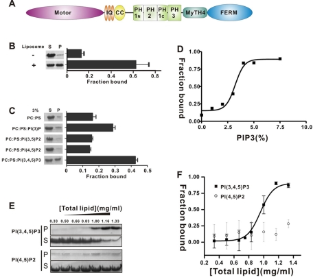
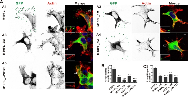
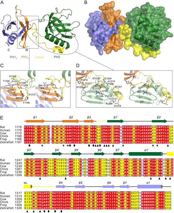
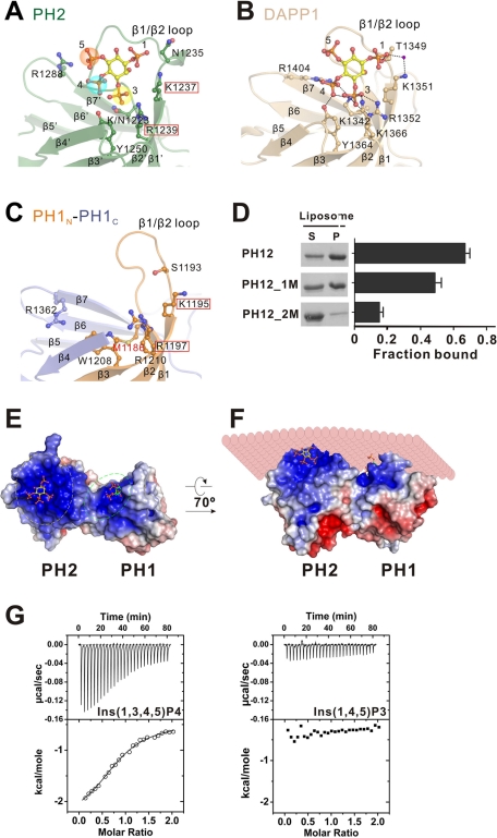
Myosin X (MyoX) is an unconventional myosin that is known to induce the formation and elongation of filopodia in many cell types. MyoX-induced filopodial induction requires the three PH domains in its tail region, although with unknown underlying molecular mechanisms. MyoX's first PH domain is split into halves by its second PH domain. We show here that the PH1(N)-PH2-PH1(C) tandem allows MyoX to bind to phosphatidylinositol (3,4,5)-triphosphate [PI(3,4,5)P(3)] with high specificity and cooperativity. We further show that PH2 is responsible for the specificity of the PI(3,4,5)P(3) interaction, whereas PH1 functions to enhance the lipid membrane-binding avidity of the tandem. The structure of the MyoX PH1(N)-PH2-PH1(C) tandem reveals that the split PH1, PH2, and the highly conserved interdomain linker sequences together form a rigid supramodule with two lipid-binding pockets positioned side by side for binding to phosphoinositide membrane bilayers with cooperativity. Finally, we demonstrate that disruption of PH2-mediated binding to PI(3,4,5)P(3) abolishes MyoX's function in inducing filopodial formation and elongation.

摘要

肌球蛋白 X(MyoX)是一种非常规的肌球蛋白,已知它能在许多细胞类型中诱导丝状伪足的形成和伸长。MyoX 诱导的丝状伪足诱导需要其尾部区域的三个 PH 结构域,但具体的分子机制尚不清楚。MyoX 的第一个 PH 结构域被其第二个 PH 结构域一分为二。我们在这里表明,PH1(N)-PH2-PH1(C)串联允许 MyoX 以高特异性和协同性结合到磷脂酰肌醇(3,4,5)-三磷酸 [PI(3,4,5)P(3)]。我们进一步表明,PH2 负责 PI(3,4,5)P(3)相互作用的特异性,而 PH1 则增强串联对脂质膜的结合亲和力。MyoX PH1(N)-PH2-PH1(C)串联的结构表明,分裂的 PH1、PH2 和高度保守的结构域间连接序列一起形成一个刚性超模块,两个脂质结合口袋并排排列,用于与磷酸肌醇膜双层协同结合。最后,我们证明了 PH2 介导的与 PI(3,4,5)P(3)结合的破坏会使 MyoX 丧失诱导丝状伪足形成和伸长的功能。

https://cdn.ncbi.nlm.nih.gov/pmc/blobs/8584/3216653/9480f4fb61b9/4268fig5.jpg
https://cdn.ncbi.nlm.nih.gov/pmc/blobs/8584/3216653/077875ab0518/4268fig1.jpg
https://cdn.ncbi.nlm.nih.gov/pmc/blobs/8584/3216653/57e671dcdbf7/4268fig2.jpg
https://cdn.ncbi.nlm.nih.gov/pmc/blobs/8584/3216653/082d4d7f97ba/4268fig3.jpg
https://cdn.ncbi.nlm.nih.gov/pmc/blobs/8584/3216653/b79f94c7b250/4268fig4.jpg
https://cdn.ncbi.nlm.nih.gov/pmc/blobs/8584/3216653/9480f4fb61b9/4268fig5.jpg

相似文献

1
Structural basis of the myosin X PH1(N)-PH2-PH1(C) tandem as a specific and acute cellular PI(3,4,5)P(3) sensor.肌球蛋白 X PH1(N)-PH2-PH1(C)串联结构作为一种特异且灵敏的细胞 PI(3,4,5)P(3)感受器
Mol Biol Cell. 2011 Nov;22(22):4268-78. doi: 10.1091/mbc.E11-04-0354. Epub 2011 Sep 30.
2
Cargo recognition mechanism of myosin X revealed by the structure of its tail MyTH4-FERM tandem in complex with the DCC P3 domain.肌球蛋白 X 的尾部 MyTH4-FERM 串联结构与 DCC P3 结构域复合物的结构揭示了其货物识别机制。
Proc Natl Acad Sci U S A. 2011 Mar 1;108(9):3572-7. doi: 10.1073/pnas.1016567108. Epub 2011 Feb 14.
3
PtdIns(3,4,5)P₃ is a regulator of myosin-X localization and filopodia formation.PtdIns(3,4,5)P₃ 是肌球蛋白-X 定位和丝状伪足形成的调节剂。
J Cell Sci. 2010 Oct 15;123(Pt 20):3525-34. doi: 10.1242/jcs.069609.
4
Myo1c binds phosphoinositides through a putative pleckstrin homology domain.肌球蛋白1c通过一个假定的普列克底物蛋白同源结构域结合磷酸肌醇。
Mol Biol Cell. 2006 Nov;17(11):4856-65. doi: 10.1091/mbc.e06-05-0449. Epub 2006 Sep 13.
5
Phospholipid-dependent regulation of the motor activity of myosin X.磷脂依赖性调节肌球蛋白 X 的运动活性。
Nat Struct Mol Biol. 2011 Jun 12;18(7):783-8. doi: 10.1038/nsmb.2065.
6
Myosin-X: a MyTH-FERM myosin at the tips of filopodia.肌球蛋白 X:丝状伪足尖端的一个 MyTH-FERM 肌球蛋白。
J Cell Sci. 2011 Nov 15;124(Pt 22):3733-41. doi: 10.1242/jcs.023549.
7
Localization of myosin 1b to actin protrusions requires phosphoinositide binding.肌球蛋白 1b 定位于肌动蛋白突起需要磷酸肌醇结合。
J Biol Chem. 2010 Sep 3;285(36):27686-93. doi: 10.1074/jbc.M109.087270. Epub 2010 Jul 7.
8
MyTH4-FERM myosins have an ancient and conserved role in filopod formation.MyTH4-FERM肌球蛋白在丝状伪足形成中具有古老且保守的作用。
Proc Natl Acad Sci U S A. 2016 Dec 13;113(50):E8059-E8068. doi: 10.1073/pnas.1615392113. Epub 2016 Nov 23.
9
Myo10 tail is crucial for promoting long filopodia.肌球蛋白 10 的尾部对于促进长丝状伪足的形成至关重要。
J Biol Chem. 2024 Jan;300(1):105523. doi: 10.1016/j.jbc.2023.105523. Epub 2023 Dec 2.
10
Association of frabin with specific actin and membrane structures.弗拉宾与特定肌动蛋白和膜结构的关联。
Genes Cells. 2002 Apr;7(4):413-20. doi: 10.1046/j.1365-2443.2002.00524.x.

引用本文的文献

1
Actin from within - how nuclear myosins and actin regulate nuclear architecture and mechanics.来自内部的肌动蛋白——核肌球蛋白和肌动蛋白如何调节核结构与力学
J Cell Sci. 2025 Feb 1;138(3). doi: 10.1242/jcs.263550. Epub 2025 Feb 10.
2
Hundreds of myosin 10s are pushed to the tips of filopodia and could cause traffic jams on actin.数以百计的肌球蛋白 10 被推到纤毛的顶端,可能导致肌动蛋白的交通堵塞。
Elife. 2024 Oct 31;12:RP90603. doi: 10.7554/eLife.90603.
3
Previously reported CCDC26 risk variant and novel germline variants in GALNT13, AR, and MYO10 associated with familial glioma in Finland.

本文引用的文献

1
Phospholipid-dependent regulation of the motor activity of myosin X.磷脂依赖性调节肌球蛋白 X 的运动活性。
Nat Struct Mol Biol. 2011 Jun 12;18(7):783-8. doi: 10.1038/nsmb.2065.
2
Structural basis of cargo recognition by the myosin-X MyTH4-FERM domain.肌球蛋白-X 的 MyTH4-FERM 结构域识别货物的结构基础。
EMBO J. 2011 Jun 3;30(13):2734-47. doi: 10.1038/emboj.2011.177.
3
Cargo recognition mechanism of myosin X revealed by the structure of its tail MyTH4-FERM tandem in complex with the DCC P3 domain.肌球蛋白 X 的尾部 MyTH4-FERM 串联结构与 DCC P3 结构域复合物的结构揭示了其货物识别机制。
先前报道的 CCDC26 风险变异和 GALNT13、AR 和 MYO10 中的新型种系变异与芬兰家族性脑胶质瘤有关。
Sci Rep. 2024 May 21;14(1):11562. doi: 10.1038/s41598-024-62296-5.
4
Hundreds of myosin 10s are pushed to the tips of filopodia and could cause traffic jams on actin.数百个肌球蛋白10被推到丝状伪足的尖端,可能会在肌动蛋白上造成交通堵塞。
bioRxiv. 2024 Aug 15:2023.06.26.546598. doi: 10.1101/2023.06.26.546598.
5
An update on genetically encoded lipid biosensors.遗传编码脂质生物传感器的最新进展。
Mol Biol Cell. 2022 May 1;33(5). doi: 10.1091/mbc.E21-07-0363.
6
Detection of Plasma Membrane Phosphoinositide Dynamics Using Genetically Encoded Fluorescent Protein Probes.利用基因编码荧光蛋白探针检测质膜磷酯酰肌醇动力学。
Methods Mol Biol. 2021;2251:73-89. doi: 10.1007/978-1-0716-1142-5_5.
7
In vitro reconstitution reveals phosphoinositides as cargo-release factors and activators of the ARF6 GAP ADAP1.体外重建揭示了磷酯酰肌醇作为货物释放因子和 ARF6 GAP ADAP1 的激活剂。
Proc Natl Acad Sci U S A. 2021 Jan 5;118(1). doi: 10.1073/pnas.2010054118. Epub 2020 Dec 18.
8
The Myosin Family of Mechanoenzymes: From Mechanisms to Therapeutic Approaches.肌球蛋白机械酶家族:从机制到治疗方法。
Annu Rev Biochem. 2020 Jun 20;89:667-693. doi: 10.1146/annurev-biochem-011520-105234. Epub 2020 Mar 13.
9
Redefining PTB domain into independently functional dual cores.将 PTB 结构域重新定义为独立功能的双核。
Biochem Biophys Res Commun. 2020 Apr 9;524(3):595-607. doi: 10.1016/j.bbrc.2020.01.114. Epub 2020 Feb 4.
10
Unconventional Myosins: How Regulation Meets Function.非传统肌球蛋白:调节与功能的交汇。
Int J Mol Sci. 2019 Dec 20;21(1):67. doi: 10.3390/ijms21010067.
Proc Natl Acad Sci U S A. 2011 Mar 1;108(9):3572-7. doi: 10.1073/pnas.1016567108. Epub 2011 Feb 14.
4
PtdIns(3,4,5)P₃ is a regulator of myosin-X localization and filopodia formation.PtdIns(3,4,5)P₃ 是肌球蛋白-X 定位和丝状伪足形成的调节剂。
J Cell Sci. 2010 Oct 15;123(Pt 20):3525-34. doi: 10.1242/jcs.069609.
5
Nerve growth factor induces axonal filopodia through localized microdomains of phosphoinositide 3-kinase activity that drive the formation of cytoskeletal precursors to filopodia.神经生长因子通过局部磷酸肌醇 3-激酶活性微域诱导轴突丝状伪足,该活性微域驱动丝状伪足形成细胞骨架前体。
J Neurosci. 2010 Sep 8;30(36):12185-97. doi: 10.1523/JNEUROSCI.1740-10.2010.
6
Myosin-X induces filopodia by multiple elongation mechanism.肌球蛋白 X 通过多种伸长机制诱导丝状伪足。
J Biol Chem. 2010 Jun 18;285(25):19605-14. doi: 10.1074/jbc.M109.093864. Epub 2010 Apr 13.
7
Myosin-X is required for cranial neural crest cell migration in Xenopus laevis.肌球蛋白-X 在非洲爪蟾颅神经嵴细胞迁移中是必需的。
Dev Dyn. 2009 Oct;238(10):2522-9. doi: 10.1002/dvdy.22077.
8
Myosin-X is critical for migratory ability of Xenopus cranial neural crest cells.肌球蛋白-X对非洲爪蟾颅神经嵴细胞的迁移能力至关重要。
Dev Biol. 2009 Nov 1;335(1):132-42. doi: 10.1016/j.ydbio.2009.08.018. Epub 2009 Aug 25.
9
Phaser crystallographic software.相位结晶学软件。
J Appl Crystallogr. 2007 Aug 1;40(Pt 4):658-674. doi: 10.1107/S0021889807021206. Epub 2007 Jul 13.
10
A novel form of motility in filopodia revealed by imaging myosin-X at the single-molecule level.通过单分子水平成像肌球蛋白-X揭示丝状伪足中一种新的运动形式。
Curr Biol. 2009 Jun 9;19(11):967-73. doi: 10.1016/j.cub.2009.03.067. Epub 2009 Apr 23.