Yan Zhi-chao, Bai Yu-jing, Tian Zhen, Hu Hai-yan, You Xiu-hua, Lin Jian-xian, Liu Shao-rui, Zhuo Ye-hong, Luo Rong-jiang
Department of Ophthalmology, The First Affiliated Hospital, Sun Yat-Sen University, Guangzhou, Guangdong, People’s Republic of China.
Mol Vis. 2011;17:2495-506. Epub 2011 Sep 27.
To investigate the efficacy, safety, and mechanisms of Sirolimus sustained delivery film on prevention of scar formation in a rabbit model of glaucoma filtration surgery.
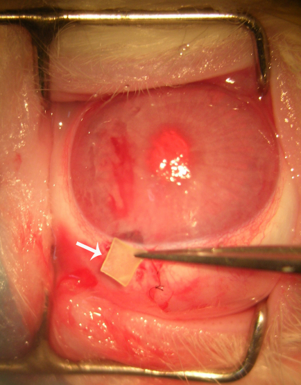
Sixty-four New Zealand white rabbits who underwent trabeculectomy in the right eye were randomly allocated to one of the four treatment regimens: Sirolimus sustained delivery film treatment group (Group A), or drug-free film treatment group (Group B), or 30 ng/ml Sirolimus-soaked sponge treatment group (Group C), or no adjunctive treatment group (Group D), and each group consists of 16 rabbits. Intraocular pressure (IOP), morphologic changes of bleb, anterior chamber flare, and corneal endothelial cell count and complications were evaluated over a 28-day period follow-up time. Aqueous humor samples were gathered from Group A, and the concentration of Sirolimus was measured regularly post-operation. Rabbits were sacrificed on the 7th, 14th, and 28th day post-operation separately, and the fibroblast hypertrophy, infiltration of inflammatory, and proliferation of new collagen fiber formation in each group were evaluated with HE and Masson staining. Proliferative cell nuclear antigen (PCNA) and fibroblast apoptosis were evaluated by immunohistochemistry and terminal deoxynucleotidyl transferasemediated dUTP nick end labeling (TUNEL) assay at the 28th day post-operation.
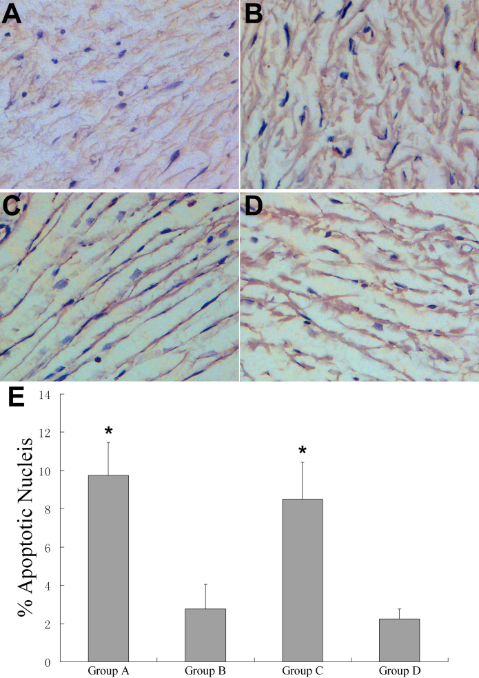
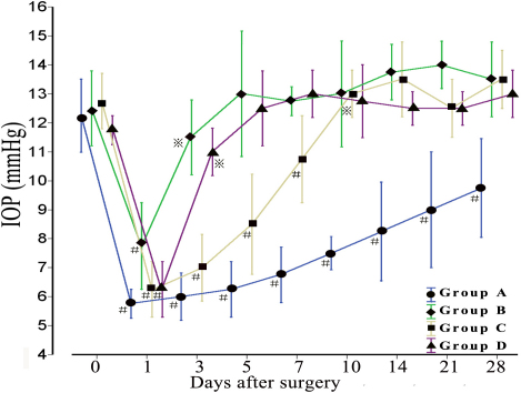
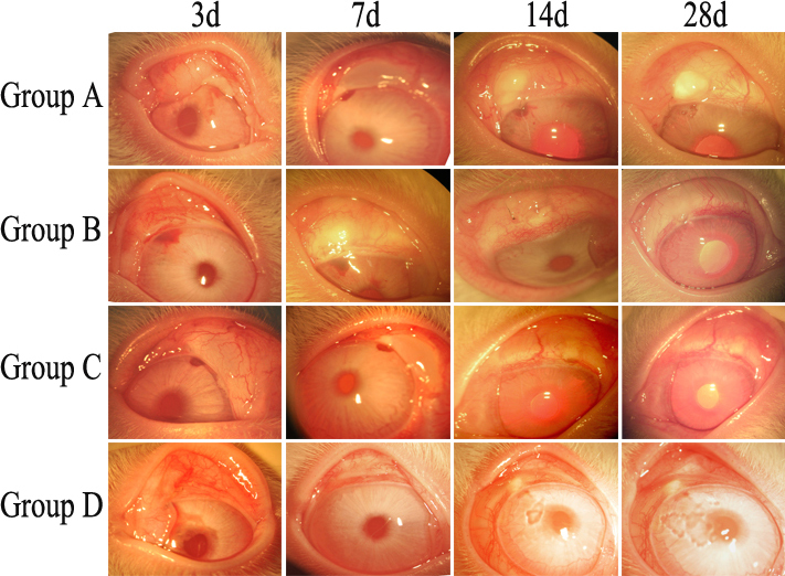
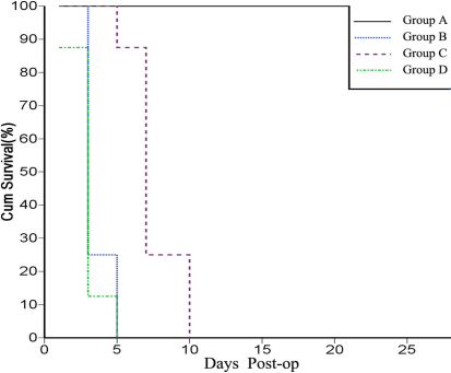
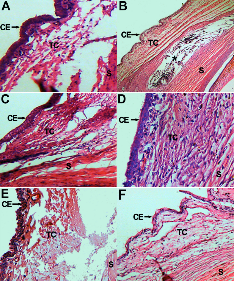
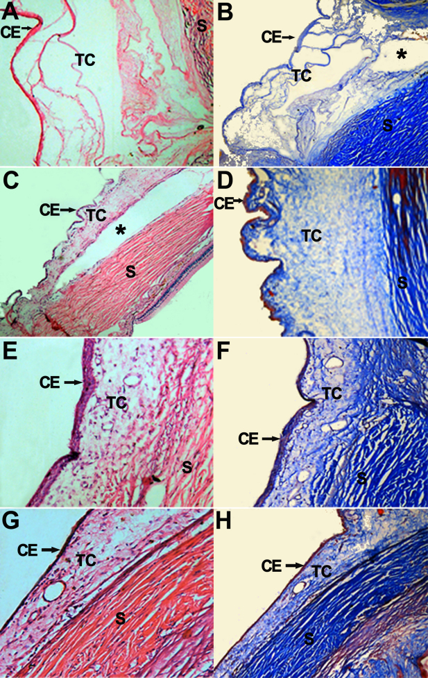
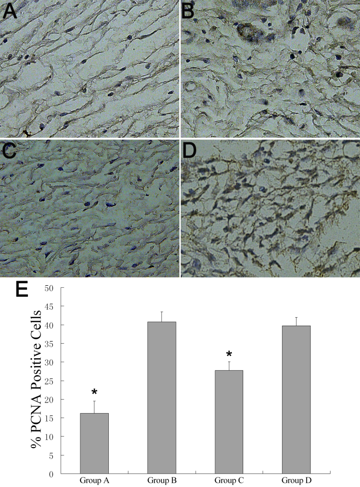
Both Sirolimus sustained delivery film (Group A) and Sirolimus alone (Group C) were well tolerated in this model, and significantly prolonged bleb survival compared with no drug treatment group (Group B and D; p<0.001). Group A had the longest bleb survival time in comparison with other groups (p<0.001). There were significant differences in IOP readings between Group A and other groups at the last follow-up (p<0.05). The concentration of Group A maintained stable for over 2 weeks, drops from (10.56 ±0.05) ng/ml at day 3 to (7.74 ±0.05) ng/ml at day 14. The number of corneal endothelial cells of Group A was not statistically significant between pre and post-operation. Histologic examination demonstrated that eyes treated with Sirolimus, especially the Sirolimus sustained delivery film, showed an obvious reduction in subconjunctival fibroblast scar tissue formation compared with no drug treatment groups, and had minimal evidence of inflammatory cell infiltration and new collagen deposition in the subconjunctiva. Immunohistochemistry assay showed that PCNA-expression was lower in the Group A (16.25±3.24%) compared to other groups (p<0.01). TUNEL assay showed a significant increase in the number of apoptotic fibroblasts around the surgical area in Group A and Group C (9.75±1.71% and 8.50±1.92%) compared to the Group B and D (p<0.01).
Sirolimus drug sustained delivery film can inhibit inflammatory cell activity, impede fibroblast proliferation activity, and induce fibroblast apoptosis in the filtration surgery sites in rabbit. The results indicate a safe and effective treatment strategy in anti-scaring treatment in glaucoma surgery.
在兔青光眼滤过手术模型中研究西罗莫司缓释膜预防瘢痕形成的疗效、安全性及机制。
64只右眼行小梁切除术的新西兰白兔被随机分为4种治疗方案之一:西罗莫司缓释膜治疗组(A组)、无药膜治疗组(B组)、30 ng/ml西罗莫司浸泡海绵治疗组(C组)或无辅助治疗组(D组),每组16只兔。在28天的随访期内评估眼压(IOP)、滤过泡形态变化、前房闪光、角膜内皮细胞计数及并发症。从A组采集房水样本,术后定期测量西罗莫司浓度。分别在术后第7天、14天和28天处死兔子,用苏木精-伊红(HE)和马松染色评估每组中纤维母细胞肥大、炎症浸润及新胶原纤维形成的增殖情况。在术后第28天通过免疫组织化学和末端脱氧核苷酸转移酶介导的dUTP缺口末端标记(TUNEL)法评估增殖细胞核抗原(PCNA)和纤维母细胞凋亡情况。
在该模型中,西罗莫司缓释膜(A组)和单独使用西罗莫司(C组)耐受性良好,与无药物治疗组(B组和D组)相比,显著延长了滤过泡存活时间(p<0.001)。与其他组相比,A组滤过泡存活时间最长(p<0.001)。在最后一次随访时,A组与其他组的眼压读数有显著差异(p<0.05)。A组浓度在2周多时间内保持稳定,从术后第3天的(10.56±0.05)ng/ml降至第14天的(7.74±0.05)ng/ml。A组术前和术后角膜内皮细胞数量无统计学差异。组织学检查表明,与无药物治疗组相比,用西罗莫司治疗的眼睛,尤其是西罗莫司缓释膜治疗的眼睛,结膜下纤维母细胞瘢痕组织形成明显减少,结膜下炎症细胞浸润和新胶原沉积的证据最少。免疫组织化学分析表明,与其他组相比,A组PCNA表达较低(16.25±3.24%)(p<0.01)。TUNEL分析表明,与B组和D组相比,A组和C组手术区域周围凋亡纤维母细胞数量显著增加(9.75±1.71%和8.50±1.92%)(p<0.01)。
西罗莫司药物缓释膜可抑制兔滤过手术部位的炎症细胞活性,阻碍纤维母细胞增殖活性,并诱导纤维母细胞凋亡。结果表明在青光眼手术抗瘢痕治疗中是一种安全有效的治疗策略。