Department of Radiology, Anzhen Hospital, Capital Medical University, Beijing, China.
J Magn Reson Imaging. 2012 Jan;35(1):72-8. doi: 10.1002/jmri.22652. Epub 2011 Oct 11.
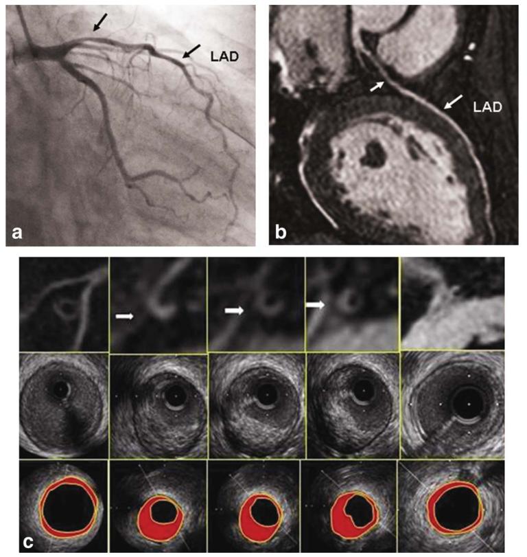
To evaluate the ability of black-blood coronary arterial wall MRI to identify the coronary artery plaque, using intravascular ultrasound (IVUS) as the golden standard.
Nineteen consecutive patients underwent IVUS and coronary artery wall MRI. Cross-sectional images were acquired on the lesion of coronary artery from the ostium to the middle segment continuously. The vessel cross-sectional area (CSA), luminal CSA, plaque burden, contrast-to-noise ratio (CNR) and signal-to-noise ratio (SNR) were measured in each slice which was then compared with the IVUS images.
Sixteen of 19 patients completed coronary artery MRA and wall imaging. 41 of 67 slices were found plaques on both IVUS and MRI; The maximal wall thickness, plaque burden, SNR, CNR in the coronary wall containing plaque were greater compared with the normal coronary wall (1.70 ± 0.51 versus 1.24 ± 0.24; 0.71 ± 0.13 versus 0.59 ± 0.12; 1.86 ± 0.41 versus 1.47 ± 0.23; 5.10 ± 2.21 versus 2.99 ± 1.17; respectively, P < 0.05). The matched MRI and IVUS showed good correlation for vessel CSA (16.77 ± 10.67 versus 16.97 ± 8.36; r = 0.79; P < 0.01), luminal CSA (5.18 ± 5.01 versus 7.13 ± 5.14; r = 0.88; P < 0.01), plaque burden (0.71 ± 0.13 versus 0.59 ± 0.15; r = 0.67; P < 0.01). in segments containing plaques, especially the luminal CSA were strongly correlated.
MRI coronary artery wall imaging can identify coronary plaque in the proximal segments. It also has the potential to assess coronary artery size.
利用血管内超声(IVUS)作为金标准,评估黑血冠状动脉壁 MRI 识别冠状动脉斑块的能力。
19 例连续患者接受了 IVUS 和冠状动脉壁 MRI 检查。在冠状动脉的病变处从开口到中段连续采集横截面图像。在每个切片上测量血管横截面积(CSA)、管腔 CSA、斑块负荷、对比噪声比(CNR)和信噪比(SNR),然后与 IVUS 图像进行比较。
19 例患者中有 16 例完成了冠状动脉 MRA 和壁成像。在 IVUS 和 MRI 上均发现 67 个切片中有 41 个斑块;含斑块的冠状动脉壁的最大壁厚度、斑块负荷、SNR、CNR 均大于正常冠状动脉壁(1.70 ± 0.51 比 1.24 ± 0.24;0.71 ± 0.13 比 0.59 ± 0.12;1.86 ± 0.41 比 1.47 ± 0.23;5.10 ± 2.21 比 2.99 ± 1.17;均 P < 0.05)。匹配的 MRI 和 IVUS 显示血管 CSA(16.77 ± 10.67 比 16.97 ± 8.36;r = 0.79;P < 0.01)、管腔 CSA(5.18 ± 5.01 比 7.13 ± 5.14;r = 0.88;P < 0.01)、斑块负荷(0.71 ± 0.13 比 0.59 ± 0.15;r = 0.67;P < 0.01)具有良好的相关性。在含有斑块的节段中,尤其是管腔 CSA 具有很强的相关性。
MRI 冠状动脉壁成像可以识别近端节段的冠状动脉斑块,也具有评估冠状动脉大小的潜力。