Department of Biology, Indiana University-Bloomington, Bloomington, Indiana, USA.
mBio. 2011 Oct 11;2(5). doi: 10.1128/mBio.00202-11. Print 2011.
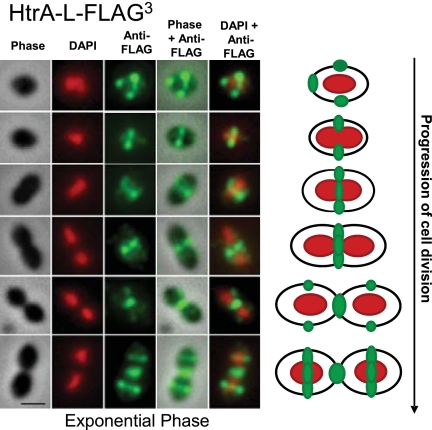
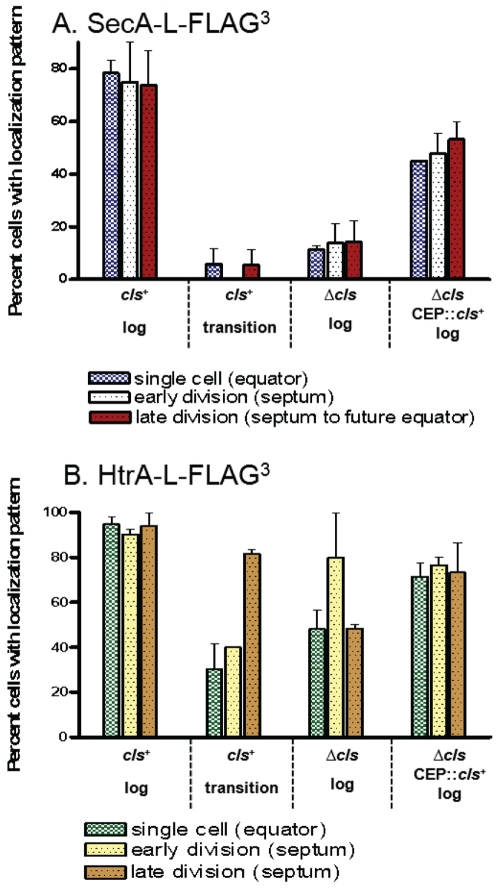
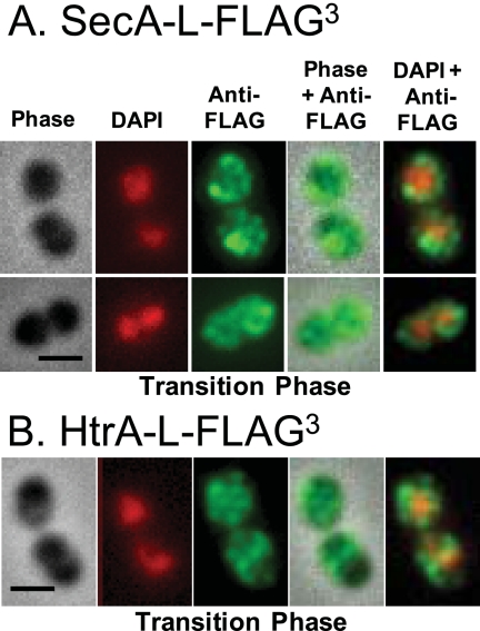
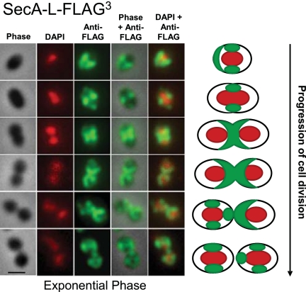
The Sec translocase pathway is the major route for protein transport across and into the cytoplasmic membrane of bacteria. Previous studies reported that the SecA translocase ATP-binding subunit and the cell surface HtrA protease/chaperone formed a single microdomain, termed "ExPortal," in some species of ellipsoidal (ovococcus) Gram-positive bacteria, including Streptococcus pyogenes. To investigate the generality of microdomain formation, we determined the distribution of SecA and SecY by immunofluorescent microscopy in Streptococcus pneumoniae (pneumococcus), which is an ovococcus species evolutionarily distant from S. pyogenes. In the majority (≥ 75%) of exponentially growing cells, S. pneumoniae SecA (SecA (Spn)) and SecY (Spn) located dynamically in cells at different stages of division. In early divisional cells, both Sec subunits concentrated at equators, which are future sites of constriction. Further along in division, SecA(Spn) and SecY(Spn) remained localized at mid-cell septa. In late divisional cells, both Sec subunits were hemispherically distributed in the regions between septa and the future equators of dividing cells. In contrast, the HtrA (Spn) homologue localized to the equators and septa of most (> 90%) dividing cells, whereas the SrtA(Spn) sortase located over the surface of cells in no discernable pattern. This dynamic pattern of Sec distribution was not perturbed by the absence of flotillin family proteins, but was largely absent in most cells in early stationary phase and in cls mutants lacking cardiolipin synthase. These results do not support the existence of an ExPortal microdomain in S. pneumoniae. Instead, the localization of the pneumococcal Sec translocase depends on the stage of cell division and anionic phospholipid content.
Two patterns of Sec translocase distribution, an ExPortal microdomain in certain ovococcus-shaped species like Streptococcus pyogenes and a spiral pattern in rod-shaped species like Bacillus subtilis, have been reported for Gram-positive bacteria. This study provides evidence for a third pattern of Sec localization in the ovococcus human pathogen Streptococcus pneumoniae. The SecA motor and SecY channel subunits of the Sec translocase localize dynamically to different places in the mid-cell region during the division cycle of exponentially growing, but not stationary-phase, S. pneumoniae. Unexpectedly, the S. pneumoniae HtrA (HtrA(Spn)) protease/chaperone principally localizes to cell equators and division septa. The coincident localization of SecA(Spn), SecY (Spn), and HtrA (Spn) to regions of peptidoglycan (PG) biosynthesis in unstressed, growing cells suggests that the pneumococcal Sec translocase directs assembly of the PG biosynthesis apparatus to regions where it is needed during division and that HtrA(Spn) may play a general role in quality control of proteins exported by the Sec translocase.
Sec 易位酶途径是蛋白质穿越和进入细菌细胞质膜的主要途径。先前的研究报道,SecA 易位酶 ATP 结合亚基和细胞表面 HtrA 蛋白酶/伴侣在某些椭圆形(卵形球菌)革兰氏阳性菌中形成一个单一的微域,称为“ExPortal”,包括酿脓链球菌。为了研究微域形成的普遍性,我们通过免疫荧光显微镜确定了肺炎链球菌(肺炎球菌)中 SecA 和 SecY 的分布,肺炎球菌是与酿脓链球菌进化上相距较远的卵形球菌种。在大多数(≥75%)指数生长期细胞中,肺炎链球菌 SecA(SecA(Spn))和 SecY(Spn)在处于不同分裂阶段的细胞中动态定位。在早期分裂细胞中,两个 Sec 亚基都集中在赤道,赤道是未来收缩的部位。进一步沿着分裂,SecA(Spn)和 SecY(Spn)仍然定位于细胞的中部隔膜。在晚期分裂细胞中,两个 Sec 亚基在隔膜之间的区域和正在分裂的细胞的未来赤道呈半球状分布。相比之下,大多数(>90%)分裂细胞中 HtrA(Spn)同源物定位于赤道和隔膜,而 SrtA(Spn)连接酶则以无明显模式分布在细胞表面。这种 Sec 分布的动态模式不受 flotillin 家族蛋白缺失的影响,但在早期静止期的大多数细胞中以及缺乏心磷脂合酶的 cls 突变体中,这种模式基本不存在。这些结果不支持肺炎球菌中存在 ExPortal 微域。相反,肺炎球菌 Sec 易位酶的定位取决于细胞分裂的阶段和阴离子磷脂的含量。
革兰氏阳性菌中已经报道了两种 Sec 易位酶分布模式,某些卵形球菌样链球菌(如酿脓链球菌)中的 ExPortal 微域和棒状杆菌(如枯草芽孢杆菌)中的螺旋模式。本研究为卵形球菌人类病原体肺炎链球菌提供了第三种 Sec 定位模式的证据。在指数生长期而不是静止期的肺炎链球菌中,Sec 易位酶的 SecA 马达和 SecY 通道亚基在细胞分裂周期中动态定位到细胞中部区域的不同位置。出乎意料的是,肺炎球菌 HtrA(HtrA(Spn))蛋白酶/伴侣主要定位于细胞赤道和分裂隔膜。在未受应激的生长细胞中,SecA(Spn)、SecY(Spn)和 HtrA(Spn)与肽聚糖(PG)生物合成区域的同时定位表明,肺炎链球菌 Sec 易位酶将 PG 生物合成装置定向到细胞分裂过程中需要的区域,并且 HtrA(Spn)可能在由 Sec 易位酶输出的蛋白质的质量控制中发挥一般作用。