Medical Biotechnology Knowledge Centre, VTT Technical Research Centre of Finland, Turku, Finland.
Oncogene. 2012 Apr 19;31(16):2075-89. doi: 10.1038/onc.2011.396. Epub 2011 Sep 26.
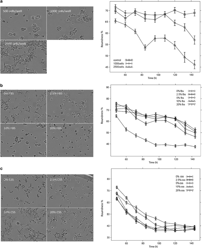
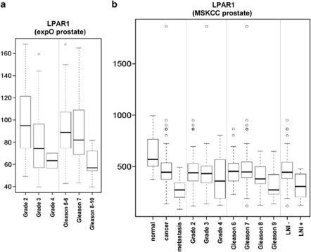
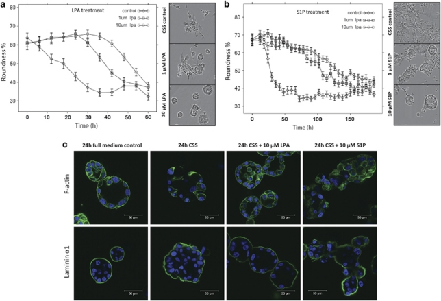
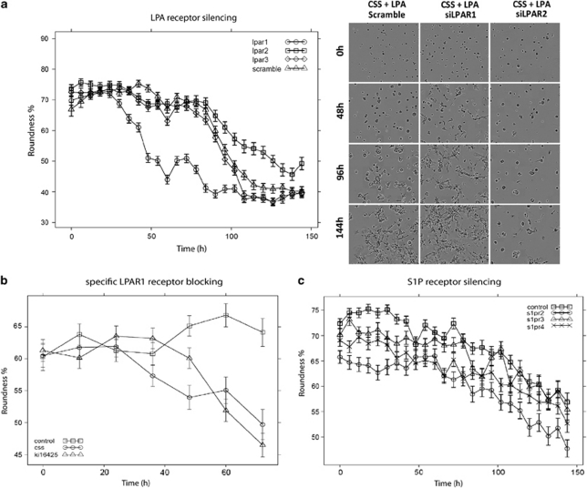
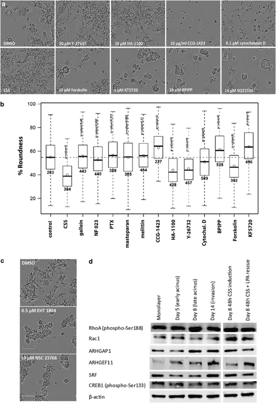
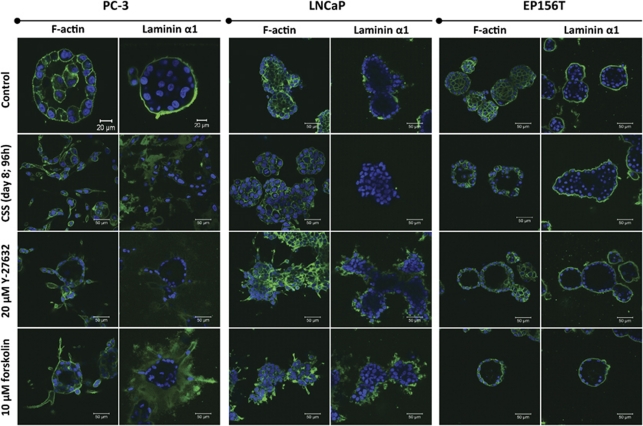
Normal prostate and some malignant prostate cancer (PrCa) cell lines undergo acinar differentiation and form spheroids in three-dimensional (3-D) organotypic culture. Acini formed by PC-3 and PC-3M, less pronounced also in other PrCa cell lines, spontaneously undergo an invasive switch, leading to the disintegration of epithelial structures and the basal lamina, and formation of invadopodia. This demonstrates the highly dynamic nature of epithelial plasticity, balancing epithelial-to-mesenchymal transition against metastable acinar differentiation. This study assessed the role of lipid metabolites on epithelial maturation. PC-3 cells completely failed to form acinar structures in delipidated serum. Adding back lysophosphatidic acid (LPA) and sphingosine-1-phosphate (S1P) rescued acinar morphogenesis and repressed invasion effectively. Blocking LPA receptor 1 (LPAR1) functions by siRNA (small interference RNA) or the specific LPAR1 inhibitor Ki16425 promoted invasion, while silencing of other G-protein-coupled receptors responsive to LPA or S1P mainly caused growth arrest or had no effects. The G-proteins Gα(12/13) and Gα(i) were identified as key mediators of LPA signalling via stimulation of RhoA and Rho kinases ROCK1 and 2, activating Rac1, while inhibition of adenylate cyclase and accumulation of cAMP may be secondary. Interfering with these pathways specifically impeded epithelial polarization in transformed cells. In contrast, blocking the same pathways in non-transformed, normal cells promoted differentiation. We conclude that LPA and LPAR1 effectively promote epithelial maturation and block invasion of PrCa cells in 3-D culture. The analysis of clinical transcriptome data confirmed reduced expression of LPAR1 in a subset of PrCa's. Our study demonstrates a metastasis-suppressor function for LPAR1 and Gα(12/13) signalling, regulating cell motility and invasion versus epithelial maturation.
正常前列腺和一些恶性前列腺癌 (PrCa) 细胞系在三维 (3-D) 器官型培养中经历腺泡分化并形成球体。PC-3 和 PC-3M 形成的腺泡,在其他 PrCa 细胞系中也不太明显,会自发发生侵袭性转换,导致上皮结构和基底膜的破坏,并形成侵袭伪足。这表明上皮可塑性具有高度动态性,平衡上皮间充质转化与亚稳态腺泡分化。本研究评估了脂质代谢物对上皮成熟的作用。去脂血清中 PC-3 细胞完全无法形成腺泡结构。添加溶血磷脂酸 (LPA) 和鞘氨醇-1-磷酸 (S1P) 可有效挽救腺泡形态发生并抑制侵袭。通过 siRNA(小干扰 RNA)或特异性 LPAR1 抑制剂 Ki16425 阻断 LPAR1 功能可促进侵袭,而沉默对 LPA 或 S1P 有反应的其他 G 蛋白偶联受体主要导致生长停滞或无影响。鉴定出 G 蛋白 Gα(12/13) 和 Gα(i) 作为 LPA 信号的关键介质,通过刺激 RhoA 和 Rho 激酶 ROCK1 和 2 激活 Rac1,而抑制腺苷酸环化酶和 cAMP 积累可能是次要的。这些途径的特异性干扰可特异性阻止转化细胞中的上皮极化。相比之下,在非转化的正常细胞中阻断相同途径可促进分化。我们得出结论,LPA 和 LPAR1 在 3-D 培养中可有效促进 PrCa 细胞的上皮成熟并阻止其侵袭。对临床转录组数据的分析证实了 LPAR1 在前列腺癌亚群中的表达降低。我们的研究表明 LPAR1 和 Gα(12/13) 信号具有抑制转移的功能,调节细胞迁移和侵袭与上皮成熟。