• 文献检索
  • 文档翻译
  • 深度研究
  • 学术资讯
  • Suppr Zotero 插件Zotero 插件
  • 邀请有礼
  • 套餐&价格
  • 历史记录
应用&插件
Suppr Zotero 插件Zotero 插件浏览器插件Mac 客户端Windows 客户端微信小程序
定价
高级版会员购买积分包购买API积分包
服务
文献检索文档翻译深度研究API 文档MCP 服务
关于我们
关于 Suppr公司介绍联系我们用户协议隐私条款
关注我们

Suppr 超能文献

核心技术专利:CN118964589B侵权必究
粤ICP备2023148730 号-1Suppr @ 2026

文献检索

告别复杂PubMed语法,用中文像聊天一样搜索,搜遍4000万医学文献。AI智能推荐,让科研检索更轻松。

立即免费搜索

文件翻译

保留排版,准确专业,支持PDF/Word/PPT等文件格式,支持 12+语言互译。

免费翻译文档

深度研究

AI帮你快速写综述,25分钟生成高质量综述,智能提取关键信息,辅助科研写作。

立即免费体验

TNFα 诱导的坏死超过凋亡的发生率取决于 TAK1-RIP1 的相互作用。

The prevalence of TNFα-induced necrosis over apoptosis is determined by TAK1-RIP1 interplay.

机构信息

Max Delbrück Center for Molecular Medicine, Berlin, Germany.

出版信息

PLoS One. 2011;6(10):e26069. doi: 10.1371/journal.pone.0026069. Epub 2011 Oct 10.

DOI:10.1371/journal.pone.0026069
PMID:22016814
原文链接:https://pmc.ncbi.nlm.nih.gov/articles/PMC3189922/
Abstract

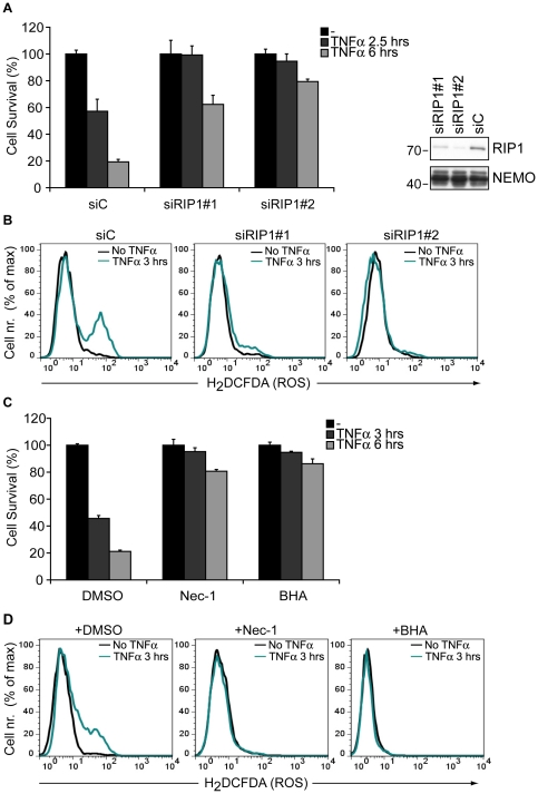
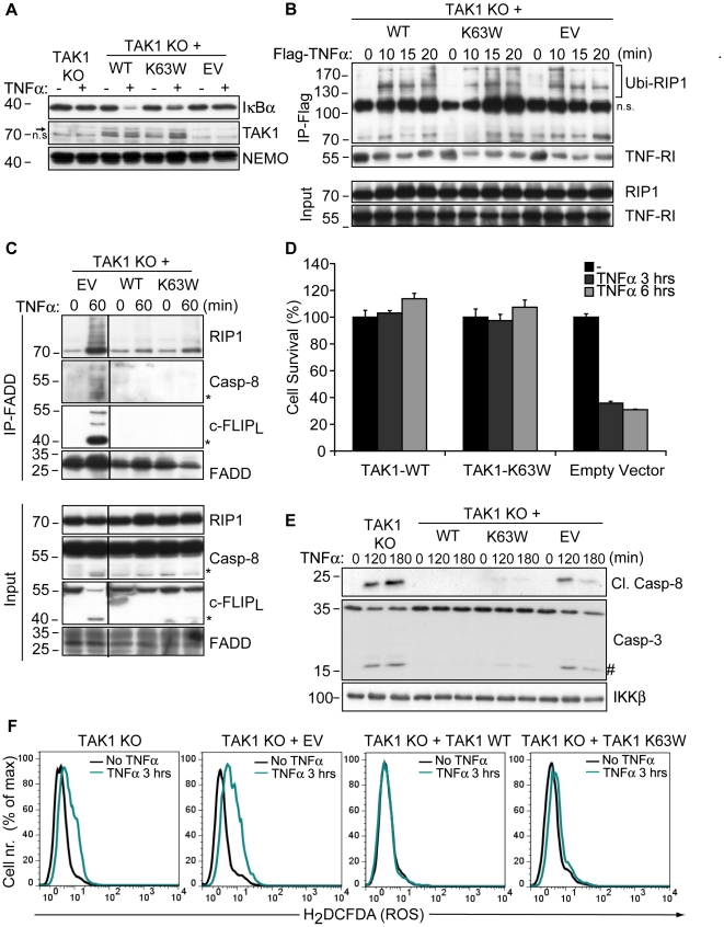
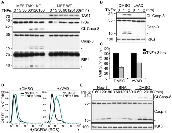
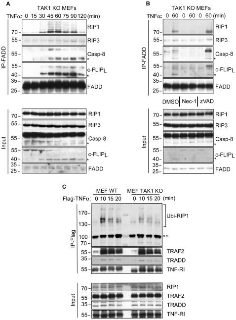
Death receptor-induced programmed necrosis is regarded as a secondary death mechanism dominating only in cells that cannot properly induce caspase-dependent apoptosis. Here, we show that in cells lacking TGFβ-activated Kinase-1 (TAK1) expression, catalytically active Receptor Interacting Protein 1 (RIP1)-dependent programmed necrosis overrides apoptotic processes following Tumor Necrosis Factor-α (TNFα) stimulation and results in rapid cell death. Importantly, the activation of the caspase cascade and caspase-8-mediated RIP1 cleavage in TNFα-stimulated TAK1 deficient cells is not sufficient to prevent RIP1-dependent necrosome formation and subsequent programmed necrosis. Our results demonstrate that TAK1 acts independently of its kinase activity to prevent the premature dissociation of ubiquitinated-RIP1 from TNFα-stimulated TNF-receptor I and also to inhibit the formation of TNFα-induced necrosome complex consisting of RIP1, RIP3, FADD, caspase-8 and cFLIP(L). The surprising prevalence of catalytically active RIP1-dependent programmed necrosis over apoptosis despite ongoing caspase activity implicates a complex regulatory mechanism governing the decision between both cell death pathways following death receptor stimulation.

摘要

死亡受体诱导的程序性细胞坏死被认为是一种次要的死亡机制,仅在不能正确诱导半胱天冬酶依赖性细胞凋亡的细胞中起作用。在这里,我们表明在缺乏 TGFβ 激活激酶 1(TAK1)表达的细胞中,催化活性的受体相互作用蛋白 1(RIP1)依赖性程序性细胞坏死会在肿瘤坏死因子-α(TNFα)刺激后取代凋亡过程,并导致快速细胞死亡。重要的是,在 TNFα 刺激的 TAK1 缺陷细胞中,胱天蛋白酶级联的激活和胱天蛋白酶-8 介导的 RIP1 裂解不足以防止 RIP1 依赖性坏死体形成和随后的程序性细胞坏死。我们的研究结果表明,TAK1 独立于其激酶活性发挥作用,以防止泛素化的 RIP1 从 TNFα 刺激的 TNF 受体 I 过早解离,并抑制由 RIP1、RIP3、FADD、胱天蛋白酶-8 和 cFLIP(L)组成的 TNFα 诱导的坏死体复合物的形成。尽管存在持续的胱天蛋白酶活性,但催化活性的 RIP1 依赖性程序性细胞坏死对细胞凋亡的普遍性,暗示了在死亡受体刺激后,两种细胞死亡途径之间的决策存在复杂的调控机制。

https://cdn.ncbi.nlm.nih.gov/pmc/blobs/5f45/3189922/efdffdc88308/pone.0026069.g004.jpg
https://cdn.ncbi.nlm.nih.gov/pmc/blobs/5f45/3189922/90b68d18c756/pone.0026069.g001.jpg
https://cdn.ncbi.nlm.nih.gov/pmc/blobs/5f45/3189922/ea67ca5269ce/pone.0026069.g002.jpg
https://cdn.ncbi.nlm.nih.gov/pmc/blobs/5f45/3189922/f495f82772af/pone.0026069.g003.jpg
https://cdn.ncbi.nlm.nih.gov/pmc/blobs/5f45/3189922/efdffdc88308/pone.0026069.g004.jpg

相似文献

1
The prevalence of TNFα-induced necrosis over apoptosis is determined by TAK1-RIP1 interplay.TNFα 诱导的坏死超过凋亡的发生率取决于 TAK1-RIP1 的相互作用。
PLoS One. 2011;6(10):e26069. doi: 10.1371/journal.pone.0026069. Epub 2011 Oct 10.
2
CYLD deubiquitinates RIP1 in the TNFα-induced necrosome to facilitate kinase activation and programmed necrosis.CYLD 在 TNFα 诱导的坏死体中去泛素化 RIP1,以促进激酶激活和程序性细胞坏死。
PLoS One. 2013 Oct 2;8(10):e76841. doi: 10.1371/journal.pone.0076841. eCollection 2013.
3
cIAP1 and TAK1 protect cells from TNF-induced necrosis by preventing RIP1/RIP3-dependent reactive oxygen species production.cIAP1 和 TAK1 通过防止 RIP1/RIP3 依赖性活性氧物种的产生来保护细胞免受 TNF 诱导的坏死。
Cell Death Differ. 2011 Apr;18(4):656-65. doi: 10.1038/cdd.2010.138. Epub 2010 Nov 5.
4
The kinase activity of Rip1 is not required for tumor necrosis factor-alpha-induced IkappaB kinase or p38 MAP kinase activation or for the ubiquitination of Rip1 by Traf2.Rip1的激酶活性对于肿瘤坏死因子-α诱导的IκB激酶或p38丝裂原活化蛋白激酶激活,或对于Traf2介导的Rip1泛素化而言并非必需。
J Biol Chem. 2004 Aug 6;279(32):33185-91. doi: 10.1074/jbc.M404206200. Epub 2004 Jun 1.
5
Cellular IAP proteins and LUBAC differentially regulate necrosome-associated RIP1 ubiquitination.细胞凋亡抑制蛋白(IAP)家族蛋白和线性泛素链组装复合体(LUBAC)对坏死小体相关的受体相互作用蛋白1(RIP1)泛素化有着不同的调控作用。
Cell Death Dis. 2015 Jun 25;6(6):e1800. doi: 10.1038/cddis.2015.158.
6
TAK1 regulates caspase 8 activation and necroptotic signaling via multiple cell death checkpoints.TAK1通过多个细胞死亡检查点调节半胱天冬酶8的激活和坏死性凋亡信号传导。
Cell Death Dis. 2016 Sep 29;7(9):e2381. doi: 10.1038/cddis.2016.294.
7
Smac mimetic triggers necroptosis in pancreatic carcinoma cells when caspase activation is blocked.当半胱天冬酶激活被阻断时,Smac模拟物可触发胰腺癌细胞发生坏死性凋亡。
Cancer Lett. 2016 Sep 28;380(1):31-8. doi: 10.1016/j.canlet.2016.05.036. Epub 2016 Jun 3.
8
Distinctive roles of receptor-interacting protein kinases 1 and 3 in caspase-independent cell death of L929.RIPK1 和 RIPK3 在 L929 细胞 caspase 非依赖性死亡中的独特作用。
Cell Biochem Funct. 2014 Jan;32(1):62-9. doi: 10.1002/cbf.2972. Epub 2013 Apr 15.
9
NIK promotes tissue destruction independently of the alternative NF-κB pathway through TNFR1/RIP1-induced apoptosis.NIK通过TNFR1/RIP1诱导的凋亡独立于替代性NF-κB途径促进组织破坏。
Cell Death Differ. 2015 Dec;22(12):2020-33. doi: 10.1038/cdd.2015.69. Epub 2015 Jun 5.
10
RIP1-dependent Bid cleavage mediates TNFα-induced but Caspase-3-independent cell death in L929 fibroblastoma cells.RIP1 依赖性的 Bid 裂解介导 L929 成纤维细胞瘤细胞中 TNFα 诱导的但不依赖于半胱天冬酶-3 的细胞死亡。
Apoptosis. 2015 Jan;20(1):92-109. doi: 10.1007/s10495-014-1058-0.

引用本文的文献

1
The Anti-Oxidative, Anti-Inflammatory, Anti-Apoptotic, and Anti-Necroptotic Role of Zinc in COVID-19 and Sepsis.锌在新冠病毒肺炎和脓毒症中的抗氧化、抗炎、抗凋亡及抗坏死性凋亡作用
Antioxidants (Basel). 2023 Oct 31;12(11):1942. doi: 10.3390/antiox12111942.
2
Tumor Necrosis Factor Family Members and Myocardial Ischemia-Reperfusion Injury: State of the Art and Therapeutic Implications.肿瘤坏死因子家族成员与心肌缺血再灌注损伤:现状与治疗意义。
Int J Mol Sci. 2023 Feb 27;24(5):4606. doi: 10.3390/ijms24054606.
3
Polymolecular botanical drug of extract (C5OSEW5050ESA) as a complementary therapy to overcome gemcitabine resistance in pancreatic cancer cells.

本文引用的文献

1
Catalytic activity of the caspase-8-FLIP(L) complex inhibits RIPK3-dependent necrosis.半胱天冬酶-8-FLIP(L) 复合物的催化活性抑制 RIPK3 依赖性坏死。
Nature. 2011 Mar 17;471(7338):363-7. doi: 10.1038/nature09852. Epub 2011 Mar 2.
2
TNFR1-induced activation of the classical NF-κB pathway.肿瘤坏死因子受体 1 诱导的经典 NF-κB 通路的激活。
FEBS J. 2011 Apr;278(6):862-76. doi: 10.1111/j.1742-4658.2011.08015.x. Epub 2011 Feb 8.
3
cIAP1 and TAK1 protect cells from TNF-induced necrosis by preventing RIP1/RIP3-dependent reactive oxygen species production.
多分子植物提取物药物(C5OSEW5050ESA)作为一种辅助疗法,用于克服胰腺癌细胞中的吉西他滨耐药性。
J Tradit Complement Med. 2022 Oct 14;13(1):39-50. doi: 10.1016/j.jtcme.2022.10.002. eCollection 2023 Jan.
4
SENP1 prevents steatohepatitis by suppressing RIPK1-driven apoptosis and inflammation.SENP1 通过抑制 RIPK1 驱动的细胞凋亡和炎症来预防脂肪性肝炎。
Nat Commun. 2022 Nov 22;13(1):7153. doi: 10.1038/s41467-022-34993-0.
5
Mechanisms of TNF-independent RIPK3-mediated cell death.TNF 非依赖型 RIPK3 介导的细胞死亡机制。
Biochem J. 2022 Oct 14;479(19):2049-2062. doi: 10.1042/BCJ20210724.
6
Cell Death Mechanisms in Cerebral Ischemia-Reperfusion Injury.脑缺血再灌注损伤中的细胞死亡机制。
Neurochem Res. 2022 Dec;47(12):3525-3542. doi: 10.1007/s11064-022-03697-8. Epub 2022 Aug 17.
7
Effect of continuous renal replacement therapy adjuvant to broad-spectrum enzyme inhibitors on the efficacy and inflammatory cytokines in patients with severe acute pancreatitis.持续肾脏替代疗法联合广谱酶抑制剂对重症急性胰腺炎患者疗效及炎症细胞因子的影响
Am J Transl Res. 2021 Jul 15;13(7):8067-8075. eCollection 2021.
8
RIPK1 can mediate apoptosis in addition to necroptosis during embryonic development.RIPK1 可以在胚胎发育过程中除了坏死性细胞凋亡外还可以介导细胞凋亡。
Cell Death Dis. 2019 Mar 13;10(3):245. doi: 10.1038/s41419-019-1490-8.
9
TAK1 mediates convergence of cellular signals for death and survival.TAK1 介导细胞死亡和存活信号的汇聚。
Apoptosis. 2019 Feb;24(1-2):3-20. doi: 10.1007/s10495-018-1490-7.
10
Model-based identification of TNFα-induced IKKβ-mediated and IκBα-mediated regulation of NFκB signal transduction as a tool to quantify the impact of drug-induced liver injury compounds.基于模型鉴定肿瘤坏死因子α诱导的IKKβ介导和IκBα介导的核因子κB信号转导调控,作为量化药物性肝损伤化合物影响的工具。
NPJ Syst Biol Appl. 2018 Jun 11;4:23. doi: 10.1038/s41540-018-0058-z. eCollection 2018.
cIAP1 和 TAK1 通过防止 RIP1/RIP3 依赖性活性氧物种的产生来保护细胞免受 TNF 诱导的坏死。
Cell Death Differ. 2011 Apr;18(4):656-65. doi: 10.1038/cdd.2010.138. Epub 2010 Nov 5.
4
Molecular mechanisms of necroptosis: an ordered cellular explosion.细胞程序性坏死的分子机制:一种有序的细胞爆炸。
Nat Rev Mol Cell Biol. 2010 Oct;11(10):700-14. doi: 10.1038/nrm2970. Epub 2010 Sep 8.
5
TAK1 suppresses a NEMO-dependent but NF-kappaB-independent pathway to liver cancer.TAK1 抑制了一种依赖于 NEMO 但不依赖于 NF-κB 的肝癌发生途径。
Cancer Cell. 2010 May 18;17(5):481-96. doi: 10.1016/j.ccr.2010.03.021.
6
The role of the kinases RIP1 and RIP3 in TNF-induced necrosis.RIP1 和 RIP3 激酶在 TNF 诱导的细胞坏死中的作用。
Sci Signal. 2010 Mar 30;3(115):re4. doi: 10.1126/scisignal.3115re4.
7
Cellular IAPs inhibit a cryptic CD95-induced cell death by limiting RIP1 kinase recruitment.细胞 IAP 可通过限制 RIP1 激酶募集来抑制隐蔽的 CD95 诱导的细胞死亡。
J Cell Biol. 2009 Dec 28;187(7):1037-54. doi: 10.1083/jcb.200904158.
8
A nuclear poly(ADP-ribose)-dependent signalosome confers DNA damage-induced IkappaB kinase activation.核多聚(ADP-核糖)依赖的信号体赋予 DNA 损伤诱导的 IkappaB 激酶的激活。
Mol Cell. 2009 Nov 13;36(3):365-78. doi: 10.1016/j.molcel.2009.09.032.
9
TRAF2 must bind to cellular inhibitors of apoptosis for tumor necrosis factor (tnf) to efficiently activate nf-{kappa}b and to prevent tnf-induced apoptosis.TRAF2 必须与细胞凋亡抑制剂结合,肿瘤坏死因子(TNF)才能有效地激活 NF-κB 并防止 TNF 诱导的细胞凋亡。
J Biol Chem. 2009 Dec 18;284(51):35906-15. doi: 10.1074/jbc.M109.072256.
10
Phosphorylation-driven assembly of the RIP1-RIP3 complex regulates programmed necrosis and virus-induced inflammation.磷酸化驱动的RIP1-RIP3复合物组装调节程序性坏死和病毒诱导的炎症。
Cell. 2009 Jun 12;137(6):1112-23. doi: 10.1016/j.cell.2009.05.037.