Department of Neurobiology, Max Planck Institute for Biophysical Chemistry, Am Fassberg 11, 37077 Göttingen, Germany.
Nature. 2011 Oct 23;479(7374):552-5. doi: 10.1038/nature10545.
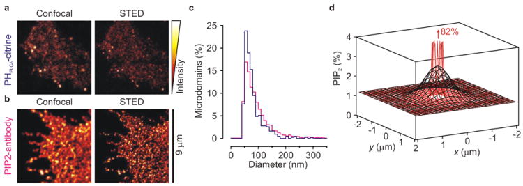
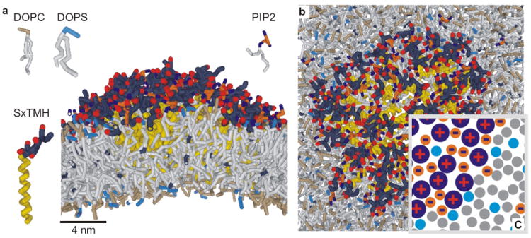
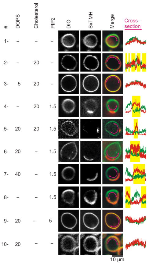
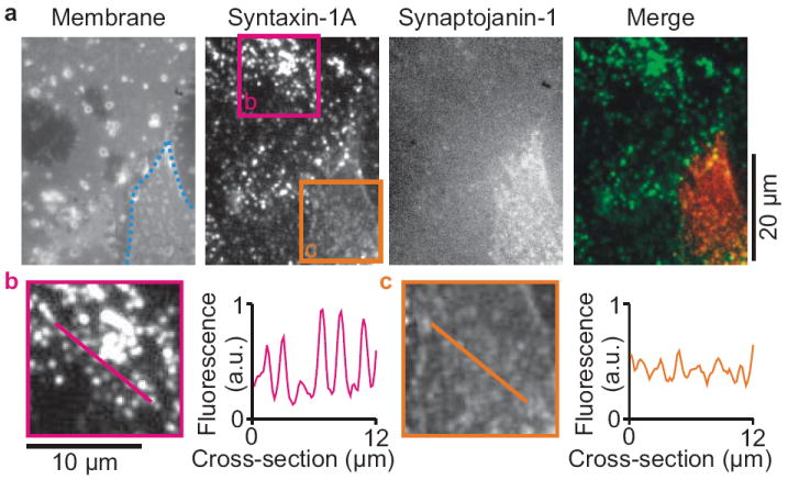
Neuronal exocytosis is catalysed by the SNAP receptor protein syntaxin-1A, which is clustered in the plasma membrane at sites where synaptic vesicles undergo exocytosis. However, how syntaxin-1A is sequestered is unknown. Here we show that syntaxin clustering is mediated by electrostatic interactions with the strongly anionic lipid phosphatidylinositol-4,5-bisphosphate (PIP2). Using super-resolution stimulated-emission depletion microscopy on the plasma membranes of PC12 cells, we found that PIP2 is the dominant inner-leaflet lipid in microdomains about 73 nanometres in size. This high accumulation of PIP2 was required for syntaxin-1A sequestering, as destruction of PIP2 by the phosphatase synaptojanin-1 reduced syntaxin-1A clustering. Furthermore, co-reconstitution of PIP2 and the carboxy-terminal part of syntaxin-1A in artificial giant unilamellar vesicles resulted in segregation of PIP2 and syntaxin-1A into distinct domains even when cholesterol was absent. Our results demonstrate that electrostatic protein-lipid interactions can result in the formation of microdomains independently of cholesterol or lipid phases.
神经元胞吐作用由 SNAP 受体蛋白突触融合蛋白 1A(syntaxin-1A)催化,该蛋白在突触小泡发生胞吐作用的部位聚集在质膜中。然而,突触融合蛋白 1A 是如何被隔离的尚不清楚。在这里,我们表明,突触融合蛋白的聚集是通过与强阴离子脂质磷脂酰肌醇-4,5-二磷酸(PIP2)的静电相互作用介导的。在 PC12 细胞的质膜上使用超分辨率受激发射损耗显微镜,我们发现 PIP2 是约 73 纳米大小的微区中的主要内层脂质。这种 PIP2 的高度积累对于突触融合蛋白 1A 的隔离是必需的,因为磷酸酶 synaptojanin-1 破坏 PIP2 会减少突触融合蛋白 1A 的聚集。此外,在人工巨大单层囊泡中共重建 PIP2 和突触融合蛋白 1A 的羧基末端部分会导致 PIP2 和突触融合蛋白 1A 即使在没有胆固醇的情况下也会分离到不同的域中。我们的结果表明,静电蛋白-脂质相互作用可以独立于胆固醇或脂质相形成微区。