Moores Cancer Center, University of California, San Diego, La Jolla, CA, USA.
J Transl Med. 2011 Oct 28;9:185. doi: 10.1186/1479-5876-9-185.
Prostate cancer metastasizes to bone in the majority of patients with advanced disease leading to painfully debilitating fractures, spinal compression and rapid decline. In addition, prostate cancer bone metastases often become resistant to standard therapies including androgen deprivation, radiation and chemotherapy. There are currently few models to elucidate mechanisms of interaction between the bone microenvironment and prostate cancer. It is, thus, essential to develop new patient-derived, orthotopic models. Here we report the development and characterization of PCSD1 (Prostate Cancer San Diego 1), a novel patient-derived intra-femoral xenograft model of prostate bone metastatic cancer that recapitulates mixed osteolytic and osteoblastic lesions.
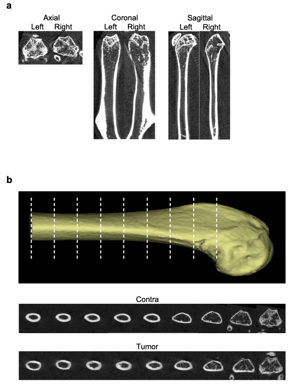
A femoral bone metastasis of prostate cancer was removed during hemiarthroplasty and transplanted into Rag2(-/-);γc(-/-) mice either intra-femorally or sub-cutaneously. Xenograft tumors that developed were analyzed for prostate cancer biomarker expression using RT-PCR and immunohistochemistry. Osteoblastic, osteolytic and mixed lesion formation was measured using micro-computed tomography (microCT).
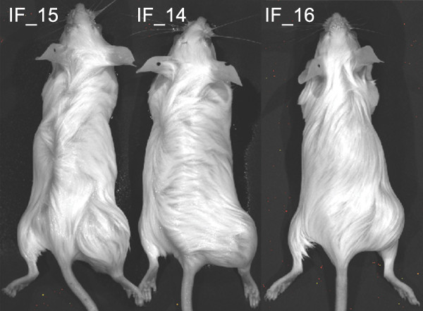
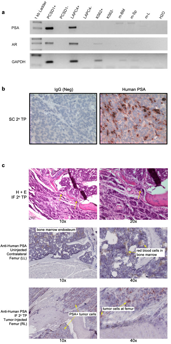
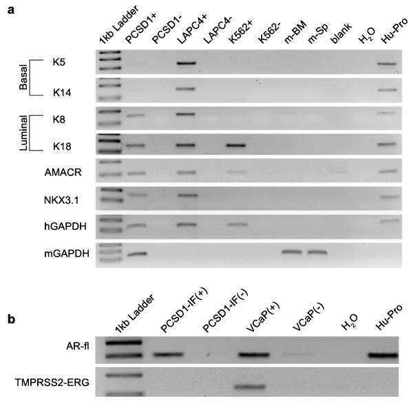
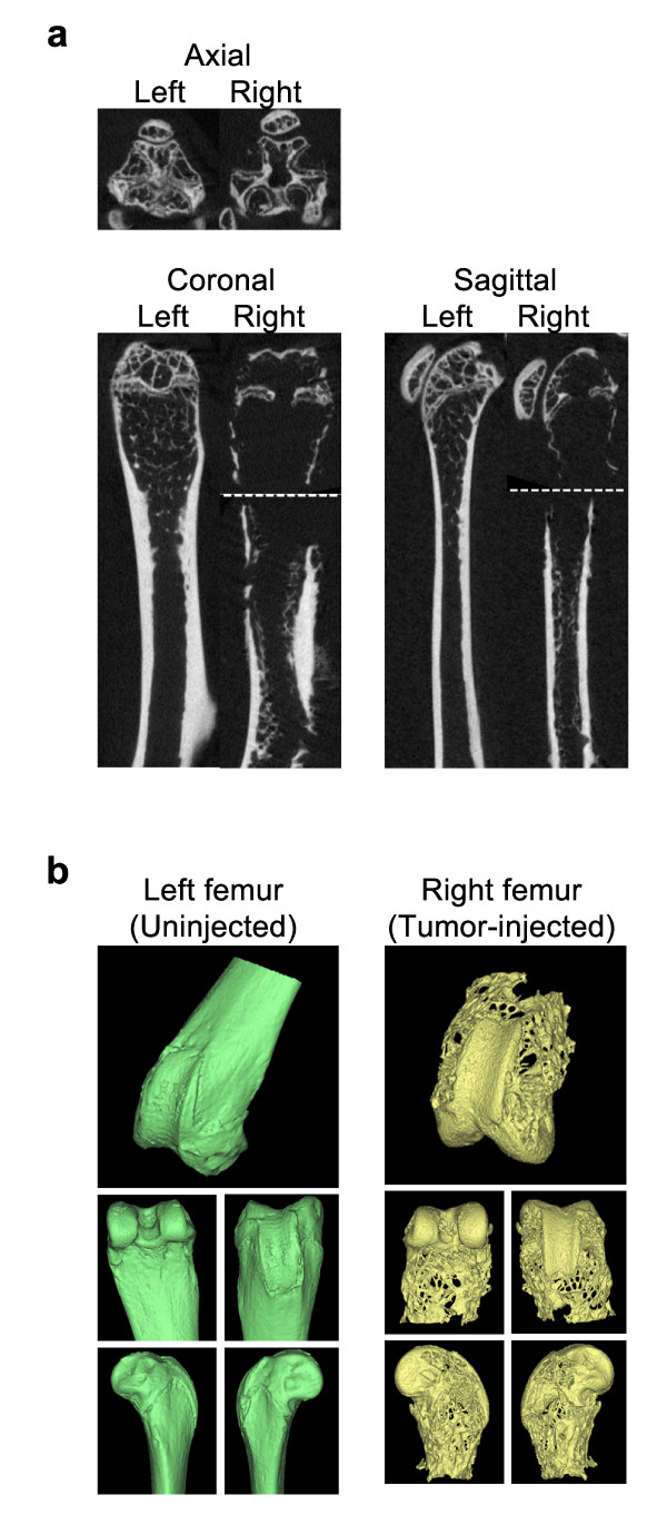
PCSD1 cells isolated directly from the patient formed tumors in all mice that were transplanted intra-femorally or sub-cutaneously into Rag2(-/-);γc(-/-) mice. Xenograft tumors expressed human prostate specific antigen (PSA) in RT-PCR and immunohistochemical analyses. PCSD1 tumors also expressed AR, NKX3.1, Keratins 8 and 18, and AMACR. Histologic and microCT analyses revealed that intra-femoral PCSD1 xenograft tumors formed mixed osteolytic and osteoblastic lesions. PCSD1 tumors have been serially passaged in mice as xenografts intra-femorally or sub-cutaneously as well as grown in culture.
PCSD1 xenografts tumors were characterized as advanced, luminal epithelial prostate cancer from a bone metastasis using RT-PCR and immunohistochemical biomarker analyses. PCSD1 intra-femoral xenografts formed mixed osteoblastic/osteolytic lesions that closely resembled the bone lesions in the patient. PCSD1 is a new primary prostate cancer bone metastasis-derived xenograft model to study metastatic disease in the bone and to develop novel therapies for inhibiting prostate cancer growth in the bone-niche.
在大多数患有晚期疾病的患者中,前列腺癌转移至骨骼,导致痛苦的虚弱性骨折、脊柱压缩和迅速恶化。此外,前列腺癌骨转移通常对包括雄激素剥夺、放疗和化疗在内的标准疗法产生耐药性。目前很少有模型可以阐明骨骼微环境与前列腺癌之间相互作用的机制。因此,开发新的患者来源的原位模型至关重要。在这里,我们报告了 PCSD1(圣地亚哥前列腺癌 1 号)的开发和特征描述,这是一种新的患者来源的股骨内原位前列腺癌骨转移模型,可重现混合溶骨性和成骨性病变。
在半髋关节置换术期间切除股骨骨转移的前列腺癌,并将其移植到 Rag2(-/-);γc(-/-) 小鼠的股骨内或皮下。使用 RT-PCR 和免疫组织化学分析来分析异种移植肿瘤中前列腺癌生物标志物的表达。使用微计算机断层扫描 (microCT) 测量成骨、溶骨和混合病变的形成。
直接从患者分离的 PCSD1 细胞在所有被移植到 Rag2(-/-);γc(-/-) 小鼠的股骨内或皮下的小鼠中形成肿瘤。异种移植肿瘤在 RT-PCR 和免疫组织化学分析中表达人前列腺特异性抗原 (PSA)。PCSD1 肿瘤还表达 AR、NKX3.1、角蛋白 8 和 18 以及 AMACR。组织学和 microCT 分析表明,股骨内 PCSD1 异种移植肿瘤形成混合溶骨性和成骨性病变。PCSD1 肿瘤已在小鼠中作为异种移植物连续传代,包括股骨内或皮下以及在培养中生长。
使用 RT-PCR 和免疫组织化学生物标志物分析,PCSD1 异种移植肿瘤被表征为来自骨转移的高级、腔上皮性前列腺癌。PCSD1 股骨内异种移植肿瘤形成混合成骨性/溶骨性病变,与患者的骨病变非常相似。PCSD1 是一种新的原发性前列腺癌骨转移源性异种移植模型,可用于研究骨骼中的转移性疾病,并开发抑制骨骼中前列腺癌生长的新疗法。