• 文献检索
  • 文档翻译
  • 深度研究
  • 学术资讯
  • Suppr Zotero 插件Zotero 插件
  • 邀请有礼
  • 套餐&价格
  • 历史记录
应用&插件
Suppr Zotero 插件Zotero 插件浏览器插件Mac 客户端Windows 客户端微信小程序
定价
高级版会员购买积分包购买API积分包
服务
文献检索文档翻译深度研究API 文档MCP 服务
关于我们
关于 Suppr公司介绍联系我们用户协议隐私条款
关注我们

Suppr 超能文献

核心技术专利:CN118964589B侵权必究
粤ICP备2023148730 号-1Suppr @ 2026

文献检索

告别复杂PubMed语法,用中文像聊天一样搜索,搜遍4000万医学文献。AI智能推荐,让科研检索更轻松。

立即免费搜索

文件翻译

保留排版,准确专业,支持PDF/Word/PPT等文件格式,支持 12+语言互译。

免费翻译文档

深度研究

AI帮你快速写综述,25分钟生成高质量综述,智能提取关键信息,辅助科研写作。

立即免费体验

炎症细胞和载脂蛋白与动脉粥样硬化进展的关联。

Association with inflammatory cells and apolipoproteins to the progression of atherosclerosis.

作者信息

Eo Hyun-Seon, Lee Kyung-Bok, Kim Ae-Kyeong, Kim Min-Hee, Kim Do-Hyung, Kim Dong-Ik

机构信息

Division of Vascular Surgery, Samsung Medical Center, Sungkyunkwan University School of Medicine, Seoul, Korea.

出版信息

J Korean Surg Soc. 2011 Apr;80(4):289-96. doi: 10.4174/jkss.2011.80.4.289. Epub 2011 Apr 12.

DOI:10.4174/jkss.2011.80.4.289
PMID:22066050
原文链接:https://pmc.ncbi.nlm.nih.gov/articles/PMC3204681/
Abstract

PURPOSE

Inflammatory cells are known to be associated with the progression of atherosclerosis and plaque rupture. However, the relation to inflammatory cells and apolipoproteins on the progression of atherosclerosis is unknown. This study was aimed at examining the different expressions of inflammatory cells and evaluate the effect of apolipoprotein (APO) C1 and APO E during the progression of atherosclerosis.

METHODS

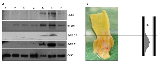
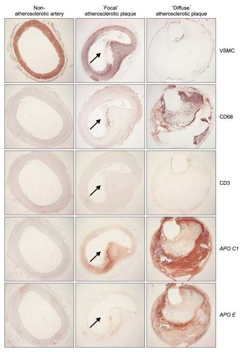
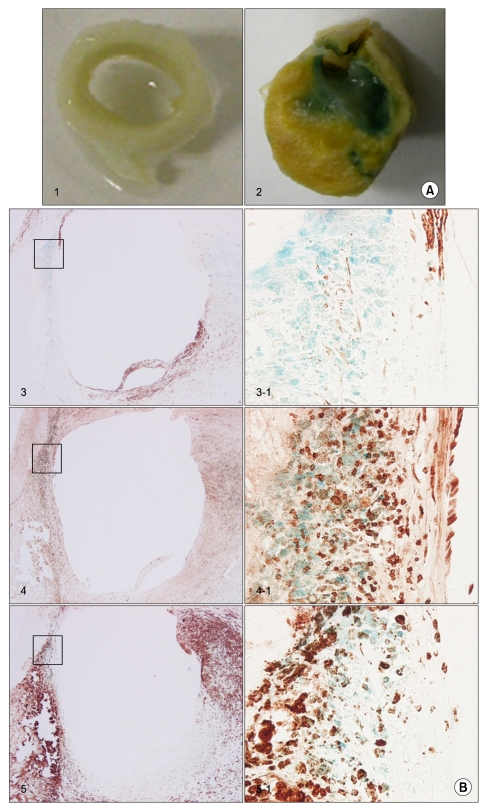
Ten atherosclerotic tissues were compared with five non-atherosclerotic tissues. The presence of vascular smooth muscle cells (VSMCs), macrophages, T-cells, APO C1, and APO E were identified by Western blotting and immunohistochemical analysis with antibodies. The senescence was analyzed by senescence-associated β-galactosidase.

RESULTS

The protein expression and senescence of macrophages, APO C1 and APO E were significantly higher in the main atherosclerotic lesion than the non-atherosclerotic lesion. A high concentration of inflammatory cells and the paucity of VSMCs were present in the shoulder area. In addition, macrophage and T-cells are expressed in the early stage of atherosclerotic development and more expanded in advanced atherosclerotic plaques. APO C1 was expressed mainly within the necrotic core, and APO E existed mostly around the necrotic core and the fibrous cap in advanced atherosclerotic plaques.

CONCLUSION

Our study indicated that the expression and the senescence of macrophage and T-cells may be closelyrelated to induction and deposition of APO C1 and APO E. This contributes to the development and progression of atherosclerotic plaque by expanding the necrotic core.

摘要

目的

已知炎症细胞与动脉粥样硬化的进展和斑块破裂相关。然而,炎症细胞和载脂蛋白与动脉粥样硬化进展之间的关系尚不清楚。本研究旨在检测炎症细胞的不同表达,并评估载脂蛋白(APO)C1和APO E在动脉粥样硬化进展过程中的作用。

方法

将10个动脉粥样硬化组织与5个非动脉粥样硬化组织进行比较。通过蛋白质免疫印迹法以及使用抗体的免疫组织化学分析来鉴定血管平滑肌细胞(VSMC)、巨噬细胞、T细胞、APO C1和APO E的存在情况。通过衰老相关β-半乳糖苷酶分析衰老情况。

结果

主要动脉粥样硬化病变中巨噬细胞、APO C1和APO E的蛋白质表达及衰老程度显著高于非动脉粥样硬化病变。肩部区域存在高浓度的炎症细胞且VSMC数量稀少。此外,巨噬细胞和T细胞在动脉粥样硬化发展的早期阶段表达,并在晚期动脉粥样硬化斑块中进一步增多。APO C1主要在坏死核心内表达,而在晚期动脉粥样硬化斑块中,APO E大多存在于坏死核心周围和纤维帽处。

结论

我们的研究表明,巨噬细胞和T细胞的表达及衰老可能与APO C1和APO E的诱导和沉积密切相关。这通过扩大坏死核心促进了动脉粥样硬化斑块的发生和发展。

https://cdn.ncbi.nlm.nih.gov/pmc/blobs/1ef4/3204681/1a02f7bc4531/jkss-80-289-g003.jpg
https://cdn.ncbi.nlm.nih.gov/pmc/blobs/1ef4/3204681/e24e58b437b8/jkss-80-289-g001.jpg
https://cdn.ncbi.nlm.nih.gov/pmc/blobs/1ef4/3204681/61df7a043e55/jkss-80-289-g002.jpg
https://cdn.ncbi.nlm.nih.gov/pmc/blobs/1ef4/3204681/1a02f7bc4531/jkss-80-289-g003.jpg
https://cdn.ncbi.nlm.nih.gov/pmc/blobs/1ef4/3204681/e24e58b437b8/jkss-80-289-g001.jpg
https://cdn.ncbi.nlm.nih.gov/pmc/blobs/1ef4/3204681/61df7a043e55/jkss-80-289-g002.jpg
https://cdn.ncbi.nlm.nih.gov/pmc/blobs/1ef4/3204681/1a02f7bc4531/jkss-80-289-g003.jpg

相似文献

1
Association with inflammatory cells and apolipoproteins to the progression of atherosclerosis.炎症细胞和载脂蛋白与动脉粥样硬化进展的关联。
J Korean Surg Soc. 2011 Apr;80(4):289-96. doi: 10.4174/jkss.2011.80.4.289. Epub 2011 Apr 12.
2
Genetic Evidence Supports a Major Role for Akt1 in VSMCs During Atherogenesis.遗传证据支持Akt1在动脉粥样硬化形成过程中对血管平滑肌细胞起主要作用。
Circ Res. 2015 May 22;116(11):1744-52. doi: 10.1161/CIRCRESAHA.116.305895. Epub 2015 Apr 13.
3
Aldehyde dehydrogenase 2 deficiency promotes atherosclerotic plaque instability through accelerating mitochondrial ROS-mediated vascular smooth muscle cell senescence.乙醛脱氢酶 2 缺乏通过加速线粒体 ROS 介导的血管平滑肌细胞衰老促进动脉粥样硬化斑块不稳定。
Biochim Biophys Acta Mol Basis Dis. 2019 Jul 1;1865(7):1782-1792. doi: 10.1016/j.bbadis.2018.09.033. Epub 2018 Oct 11.
4
Characterization of atherosclerotic lesions in apo E3-leiden transgenic mice.载脂蛋白E3-莱顿转基因小鼠动脉粥样硬化病变的特征分析
Atherosclerosis. 1998 Jan;136(1):147-52. doi: 10.1016/s0021-9150(97)00196-2.
5
Apolipoprotein E localization in human coronary atherosclerotic plaques by in situ hybridization and immunohistochemistry and comparison with lipoprotein lipase.通过原位杂交和免疫组织化学法检测载脂蛋白E在人冠状动脉粥样硬化斑块中的定位及其与脂蛋白脂肪酶的比较
Am J Pathol. 1994 Mar;144(3):538-48.
6
EphA2 Expression Regulates Inflammation and Fibroproliferative Remodeling in Atherosclerosis.EphA2表达调控动脉粥样硬化中的炎症和纤维增生性重塑。
Circulation. 2017 Aug 8;136(6):566-582. doi: 10.1161/CIRCULATIONAHA.116.026644. Epub 2017 May 9.
7
Vascular smooth muscle cells undergo telomere-based senescence in human atherosclerosis: effects of telomerase and oxidative stress.在人类动脉粥样硬化中,血管平滑肌细胞会经历基于端粒的衰老:端粒酶和氧化应激的影响。
Circ Res. 2006 Jul 21;99(2):156-64. doi: 10.1161/01.RES.0000233315.38086.bc. Epub 2006 Jun 22.
8
Vascular smooth muscle cell death, autophagy and senescence in atherosclerosis.动脉粥样硬化中的血管平滑肌细胞死亡、自噬和衰老。
Cardiovasc Res. 2018 Mar 15;114(4):622-634. doi: 10.1093/cvr/cvy007.
9
Targeting Coagulation Factor Xa Promotes Regression of Advanced Atherosclerosis in Apolipoprotein-E Deficient Mice.靶向凝血因子 Xa 可促进载脂蛋白 E 缺陷小鼠晚期动脉粥样硬化的消退。
Sci Rep. 2019 Mar 7;9(1):3909. doi: 10.1038/s41598-019-40602-w.
10
Research on the correlation between activating transcription factor 3 expression in the human coronary artery and atherosclerotic plaque stability.研究人冠状动脉中激活转录因子 3 的表达与动脉粥样硬化斑块稳定性的相关性。
BMC Cardiovasc Disord. 2021 Jul 28;21(1):356. doi: 10.1186/s12872-021-02161-9.

引用本文的文献

1
Apolipoprotein C1: Its Pleiotropic Effects in Lipid Metabolism and Beyond.载脂蛋白 C1:其在脂代谢及其他方面的多效性作用。
Int J Mol Sci. 2019 Nov 26;20(23):5939. doi: 10.3390/ijms20235939.

本文引用的文献

1
Apolipoprotein C1 and apolipoprotein E are differentially expressed in atheroma of the carotid and femoral artery.载脂蛋白C1和载脂蛋白E在颈动脉和股动脉粥样硬化斑块中存在差异表达。
J Surg Res. 2008 Jan;144(1):132-7. doi: 10.1016/j.jss.2007.02.009. Epub 2007 Oct 22.
2
Defining the role of vascular smooth muscle cell apoptosis in atherosclerosis.界定血管平滑肌细胞凋亡在动脉粥样硬化中的作用。
Cell Cycle. 2006 Oct;5(20):2329-31. doi: 10.4161/cc.5.20.3383. Epub 2006 Oct 16.
3
Short telomeres protect from diet-induced atherosclerosis in apolipoprotein E-null mice.
短端粒可保护载脂蛋白E基因敲除小鼠免受饮食诱导的动脉粥样硬化。
FASEB J. 2004 Feb;18(2):418-20. doi: 10.1096/fj.03-0710fje. Epub 2003 Dec 19.
4
Regulation of smooth muscle cell growth, function and death in vitro by activated mast cells--a potential mechanism for the weakening and rupture of atherosclerotic plaques.活化肥大细胞对体外平滑肌细胞生长、功能及死亡的调节——动脉粥样硬化斑块变薄和破裂的一种潜在机制
Biochem Pharmacol. 2003 Oct 15;66(8):1493-8. doi: 10.1016/s0006-2952(03)00503-3.
5
Changing concepts of atherogenesis.动脉粥样硬化形成的概念变迁
J Intern Med. 2000 Mar;247(3):349-58. doi: 10.1046/j.1365-2796.2000.00654.x.
6
Apoptosis in atherosclerosis: beneficial or detrimental?动脉粥样硬化中的细胞凋亡:有益还是有害?
Cardiovasc Res. 2000 Feb;45(3):736-46. doi: 10.1016/s0008-6363(99)00235-7.
7
Apolipoprotein A-I stimulates secretion of apolipoprotein E by foam cell macrophages.载脂蛋白A-I刺激泡沫细胞巨噬细胞分泌载脂蛋白E。
J Biol Chem. 1999 Sep 24;274(39):27925-33. doi: 10.1074/jbc.274.39.27925.
8
Serial analysis of gene expression in human monocytes and macrophages.人类单核细胞和巨噬细胞中基因表达的序列分析
Blood. 1999 Aug 1;94(3):837-44.
9
Biphasic pattern of cell turnover characterizes the progression from fatty streaks to ruptured human atherosclerotic plaques.
Cardiovasc Res. 1999 Feb;41(2):473-9. doi: 10.1016/s0008-6363(98)00311-3.
10
Apoptosis of vascular smooth muscle cells in atherosclerosis.
Atherosclerosis. 1998 May;138(1):3-9. doi: 10.1016/s0021-9150(98)00013-6.