Department of Medicine, Université de Montréal, CRCHUM, Pavilion J,A, de Sève, 1560 Sherbrooke E, Montreal, QC, H2L 4M1, Canada.
J Neuroinflammation. 2011 Nov 8;8:155. doi: 10.1186/1742-2094-8-155.
Multiple sclerosis (MS), an inflammatory disease of the central nervous system (CNS), is characterized by blood-brain barrier (BBB) disruption and massive infiltration of activated immune cells. Engagement of programmed cell death-1 (PD-1) expressed on activated T cells with its ligands (PD-L1 and PD-L2) suppresses T cell responses. We recently demonstrated in MS lesions elevated PD-L1 expression by glial cells and absence of PD-1 on many infiltrating CD8 T cells. We have now investigated whether human brain endothelial cells (HBECs), which maintain the BBB, can express PD-L1 or PD-L2 and thereby modulate T cells.
We used primary cultures of HBECs isolated from non-tumoral CNS tissue either under basal or inflamed conditions. We assessed the expression of PD-L1 and PD-L2 using qPCR and flow cytometry. Human CD8 T cells were isolated from peripheral blood of healthy donors and co-cultured with HBECs. Following co-culture with HBECs, proliferation and cytokine production by human CD8 T cells were measured by flow cytometry whereas transmigration was determined using a well established in vitro model of the BBB. The functional impact of PD-L1 and PD-L2 provided by HBECs was determined using blocking antibodies. We performed immunohistochemistry for the detection of PD-L1 or PD-L2 concurrently with caveolin-1 (a cell specific marker for endothelial cells) on post-mortem human brain tissues obtained from MS patients and normal controls.
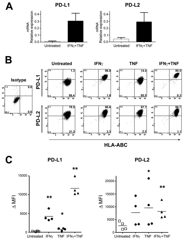
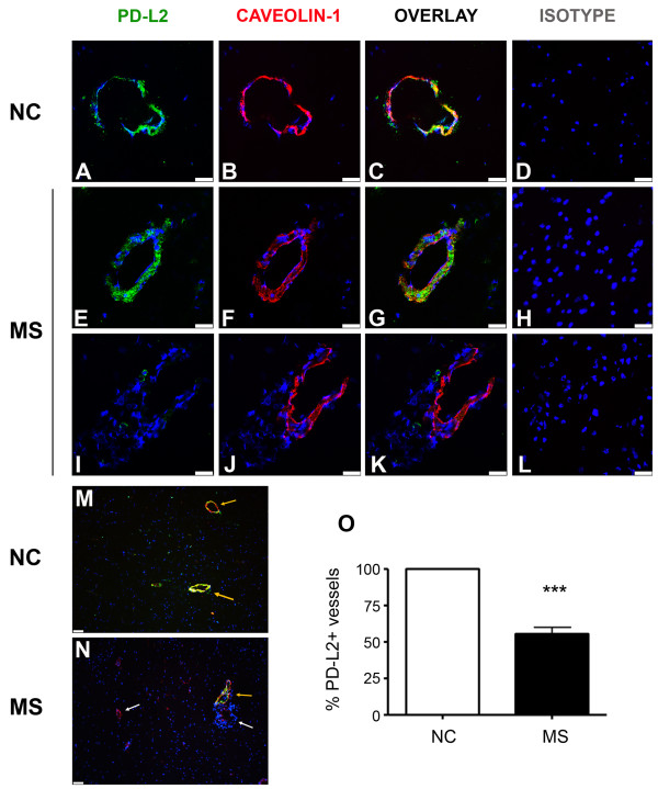
Under basal culture conditions, PD-L2 is expressed on HBECs, whilst PD-L1 is not detected. Both ligands are up-regulated under inflammatory conditions. Blocking PD-L1 and PD-L2 leads to increased transmigration and enhanced responses by human CD8 T cells in co-culture assays. Similarly, PD-L1 and PD-L2 blockade significantly increases CD4 T cell transmigration. Brain endothelium in normal tissues and MS lesions does not express detectable PD-L1; in contrast, all blood vessels in normal brain tissues are PD-L2-positive, while only about 50% express PD-L2 in MS lesions.
Our observations suggest that brain endothelial cells contribute to control T cell transmigration into the CNS and immune responses via PD-L2 expression. However, such impact is impaired in MS lesions due to downregulation of endothelium PD-L2 levels.
多发性硬化症(MS)是一种中枢神经系统(CNS)的炎症性疾病,其特征是血脑屏障(BBB)的破坏和大量活化免疫细胞的浸润。活化 T 细胞上表达的程序性细胞死亡受体-1(PD-1)与其配体(PD-L1 和 PD-L2)结合可抑制 T 细胞的反应。我们最近在 MS 病变中发现,胶质细胞表达 PD-L1 水平升高,而许多浸润的 CD8 T 细胞上缺乏 PD-1。我们现在研究了维持 BBB 的人脑微血管内皮细胞(HBEC)是否可以表达 PD-L1 或 PD-L2,从而调节 T 细胞。
我们使用从非肿瘤性 CNS 组织中分离的 HBEC 的原代培养物,在基础或炎症条件下进行研究。我们使用 qPCR 和流式细胞术评估 PD-L1 和 PD-L2 的表达。从健康供体的外周血中分离 CD8 T 细胞,并与人 HBEC 共培养。与人 HBEC 共培养后,通过流式细胞术测量人 CD8 T 细胞的增殖和细胞因子产生,而通过体外 BBB 模型确定其迁移。使用 HBEC 提供的 PD-L1 和 PD-L2 的阻断抗体来确定其功能影响。我们对从 MS 患者和正常对照中获得的死后人脑组织进行了 PD-L1 或 PD-L2 的免疫组化检测,同时检测了小窝蛋白-1(内皮细胞的一种细胞特异性标志物)。
在基础培养条件下,HBEC 表达 PD-L2,而不检测到 PD-L1。在炎症条件下,两种配体均上调。阻断 PD-L1 和 PD-L2 可导致共培养中人类 CD8 T 细胞的迁移增加和反应增强。同样,PD-L1 和 PD-L2 阻断显著增加 CD4 T 细胞的迁移。正常组织和 MS 病变中的脑内皮细胞不表达可检测到的 PD-L1;相比之下,正常脑组织中的所有血管均为 PD-L2 阳性,而在 MS 病变中只有约 50%表达 PD-L2。
我们的观察结果表明,脑微血管内皮细胞通过 PD-L2 表达来控制 T 细胞向中枢神经系统的迁移和免疫反应。但是,由于内皮细胞 PD-L2 水平下调,这种作用在 MS 病变中受损。