Neurovirology Laboratory, Department of Medicine, University of Minnesota, Minnesota, USA.
Immun Inflamm Dis. 2018 Jun;6(2):332-344. doi: 10.1002/iid3.221. Epub 2018 Mar 30.
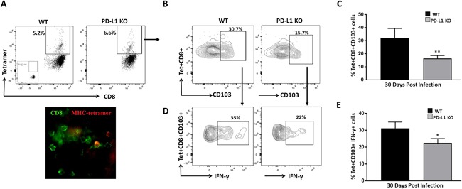
Previous work from our laboratory has demonstrated in vivo persistence of CD103 CD69 brain resident memory CD8 T-cells (bT ) following viral infection, and that the PD-1: PD-L1 pathway promotes development of these T cells within the brain. Although glial cells express low basal levels of PD-L1, its expression is upregulated upon IFN-γ-treatment, and they have been shown to modulate antiviral T-cell effector responses through the PD-1: PD-L1 pathway.
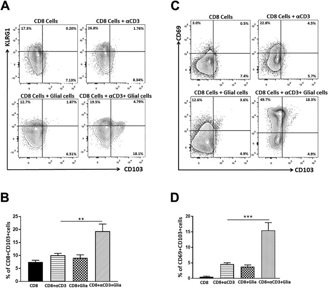
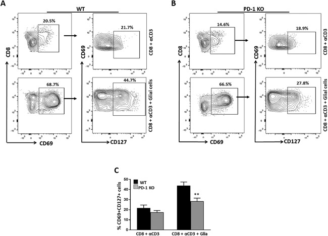
We performed flow cytometric analysis of cells from co-cultures of mixed glia and CD8 T-cells obtained from wild type mice to investigate the role of glial cells in the development of bT .
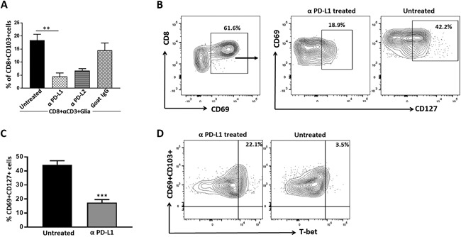
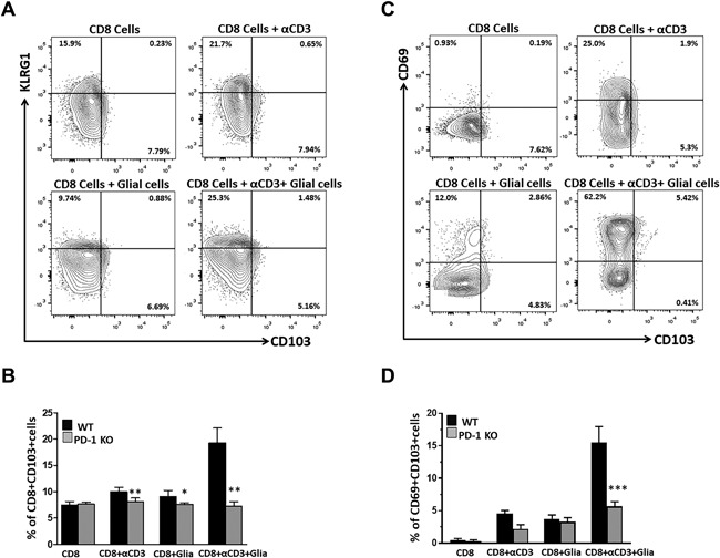
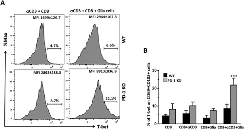
In this study, we show that interactions between reactive glia and anti-CD3 Ab-stimulated CD8 T-cells promote development of CD103 CD69 CD8 T-cells through engagement of the PD-1: PD-L1 pathway. These studies used co-cultures of primary murine glial cells obtained from WT animals along with CD8 T-cells obtained from either WT or PD-1 KO mice. We found that αCD3 Ab-stimulated CD8 T-cells from WT animals increased expression of CD103 and CD69 when co-cultured with primary murine glial cells. In contrast, significantly reduced expression of CD103 and CD69 was observed using CD8 T-cells from PD-1 KO mice. We also observed that reactive glia promoted high levels of CD127, a marker of memory precursor effector cells (MPEC), on CD69 CD8 T-cells, which promotes development of T cells. Interestingly, results obtained using T-cells from PD-1 KO animals showed significantly reduced expression of CD127 on CD69 CD8 cells. Additionally, blocking of glial PD-L1 resulted in decreased expression of CD103, along with reduced CD127 on CD69 CD8 T-cells.
Taken together, these results demonstrate a role for activated glia in promoting development of bT through the PD-1: PD-L1 pathway.
本实验室前期工作表明,在病毒感染后,CD103 CD69 脑驻留记忆 CD8 T 细胞(bT)在体内持续存在,而 PD-1:PD-L1 通路促进了这些 T 细胞在脑内的发育。尽管神经胶质细胞低表达 PD-L1,但在 IFN-γ 处理后其表达上调,并且已证明它们通过 PD-1:PD-L1 通路调节抗病毒 T 细胞效应反应。
我们对来自野生型小鼠的混合神经胶质细胞和 CD8 T 细胞共培养物中的细胞进行流式细胞术分析,以研究神经胶质细胞在 bT 发育中的作用。
在这项研究中,我们表明反应性神经胶质细胞与抗 CD3 Ab 刺激的 CD8 T 细胞之间的相互作用通过 PD-1:PD-L1 通路促进 CD103 CD69 CD8 T 细胞的发育。这些研究使用了来自 WT 动物的原代小鼠神经胶质细胞与来自 WT 或 PD-1 KO 小鼠的 CD8 T 细胞共培养。我们发现,当与原代小鼠神经胶质细胞共培养时,WT 动物的抗 CD3 Ab 刺激的 CD8 T 细胞增加了 CD103 和 CD69 的表达。相比之下,在 PD-1 KO 小鼠的 CD8 T 细胞中观察到 CD103 和 CD69 的表达显著降低。我们还观察到,反应性神经胶质细胞促进了 CD69 CD8 T 细胞上高表达 CD127,这是记忆前体细胞效应细胞(MPEC)的标志物,从而促进了 T 细胞的发育。有趣的是,使用 PD-1 KO 动物的 T 细胞获得的结果表明,CD69 CD8 细胞上 CD127 的表达显著降低。此外,阻断神经胶质细胞 PD-L1 导致 CD103 的表达减少,同时 CD69 CD8 T 细胞上 CD127 的表达减少。
综上所述,这些结果表明,激活的神经胶质细胞通过 PD-1:PD-L1 通路在促进 bT 发育中发挥作用。