Department of Biomedical Engineering, New Jersey Institute of Technology, Newark, New Jersey, United States of America.
PLoS One. 2011;6(11):e25866. doi: 10.1371/journal.pone.0025866. Epub 2011 Nov 2.
Eye movement research has traditionally studied solely saccade and/or vergence eye movements by isolating these systems within a laboratory setting. While the neural correlates of saccadic eye movements are established, few studies have quantified the functional activity of vergence eye movements using fMRI. This study mapped the neural substrates of vergence eye movements and compared them to saccades to elucidate the spatial commonality and differentiation between these systems.
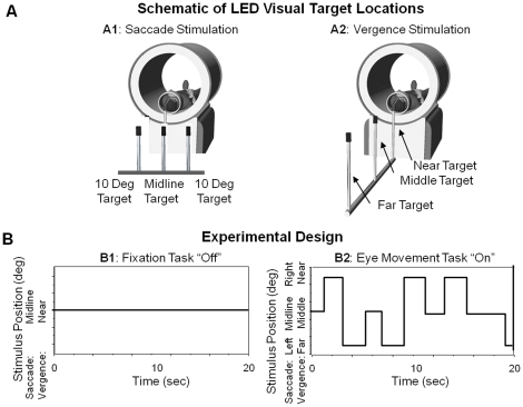
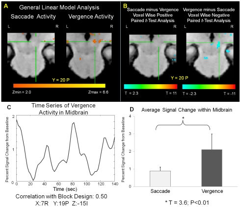
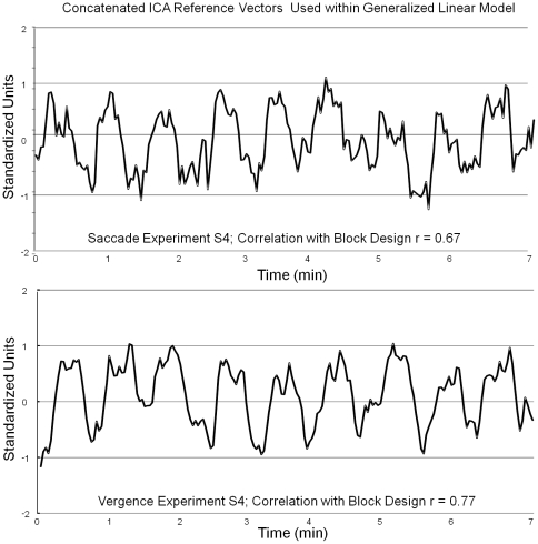
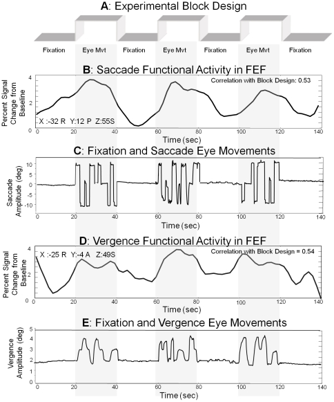
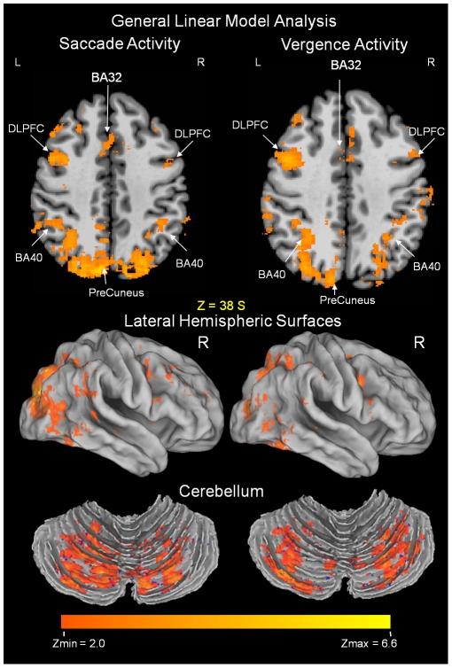
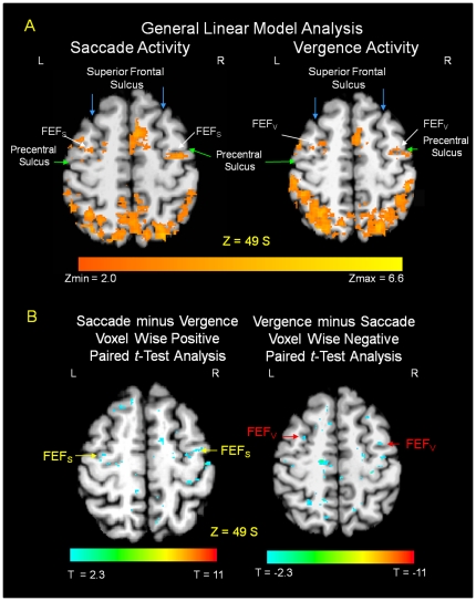
The stimulus was presented in a block design where the 'off' stimulus was a sustained fixation and the 'on' stimulus was random vergence or saccadic eye movements. Data were collected with a 3T scanner. A general linear model (GLM) was used in conjunction with cluster size to determine significantly active regions. A paired t-test of the GLM beta weight coefficients was computed between the saccade and vergence functional activities to test the hypothesis that vergence and saccadic stimulation would have spatial differentiation in addition to shared neural substrates.
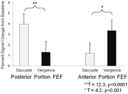
Segregated functional activation was observed within the frontal eye fields where a portion of the functional activity from the vergence task was located anterior to the saccadic functional activity (z>2.3; p<0.03). An area within the midbrain was significantly correlated with the experimental design for the vergence but not the saccade data set. Similar functional activation was observed within the following regions of interest: the supplementary eye field, dorsolateral prefrontal cortex, ventral lateral prefrontal cortex, lateral intraparietal area, cuneus, precuneus, anterior and posterior cingulates, and cerebellar vermis. The functional activity from these regions was not different between the vergence and saccade data sets assessed by analyzing the beta weights of the paired t-test (p>0.2).
Functional MRI can elucidate the differences between the vergence and saccade neural substrates within the frontal eye fields and midbrain.
眼动研究传统上仅通过在实验室环境中隔离这些系统来研究扫视和/或聚散眼动。虽然已确定了扫视眼动的神经相关性,但很少有研究使用 fMRI 量化聚散眼动的功能活动。本研究绘制了聚散眼动的神经基质,并将其与扫视进行了比较,以阐明这两个系统之间的空间共性和差异。
该刺激采用块设计呈现,“关闭”刺激是持续注视,“开启”刺激是随机聚散或扫视眼动。数据是用 3T 扫描仪收集的。使用广义线性模型 (GLM) 结合簇大小来确定显著活跃区域。通过扫视和聚散功能活动的 GLM 贝塔权重系数的配对 t 检验来计算,以检验以下假设:除了共同的神经基质外,聚散和扫视刺激还具有空间差异。
在前额叶眼区观察到功能活动的分离,其中一部分聚散任务的功能活动位于扫视功能活动的前部(z>2.3;p<0.03)。中脑内的一个区域与聚散任务的实验设计显著相关,但与扫视数据集无关。在以下感兴趣区域也观察到类似的功能激活:辅助眼区、背外侧前额叶皮质、腹外侧前额叶皮质、外侧顶内区、楔前叶、扣带回前回和后回、小脑蚓部。通过分析配对 t 检验的 beta 权重(p>0.2),评估扫视和聚散数据集之间的功能活动没有差异。
功能 MRI 可以阐明前额眼区和中脑内聚散和扫视神经基质之间的差异。