• 文献检索
  • 文档翻译
  • 深度研究
  • 学术资讯
  • Suppr Zotero 插件Zotero 插件
  • 邀请有礼
  • 套餐&价格
  • 历史记录
应用&插件
Suppr Zotero 插件Zotero 插件浏览器插件Mac 客户端Windows 客户端微信小程序
定价
高级版会员购买积分包购买API积分包
服务
文献检索文档翻译深度研究API 文档MCP 服务
关于我们
关于 Suppr公司介绍联系我们用户协议隐私条款
关注我们

Suppr 超能文献

核心技术专利:CN118964589B侵权必究
粤ICP备2023148730 号-1Suppr @ 2026

文献检索

告别复杂PubMed语法,用中文像聊天一样搜索,搜遍4000万医学文献。AI智能推荐,让科研检索更轻松。

立即免费搜索

文件翻译

保留排版,准确专业,支持PDF/Word/PPT等文件格式,支持 12+语言互译。

免费翻译文档

深度研究

AI帮你快速写综述,25分钟生成高质量综述,智能提取关键信息,辅助科研写作。

立即免费体验

虐待性头部创伤 第二部分:影像学表现。

Abusive head trauma Part II: radiological aspects.

机构信息

Section Forensic Paediatrics, Department of Forensic Medicine, Netherlands Forensic Institute, The Hague, The Netherlands.

出版信息

Eur J Pediatr. 2012 Apr;171(4):617-23. doi: 10.1007/s00431-011-1611-6. Epub 2011 Nov 15.

DOI:10.1007/s00431-011-1611-6
PMID:22080958
原文链接:https://pmc.ncbi.nlm.nih.gov/articles/PMC3306566/
Abstract

UNLABELLED

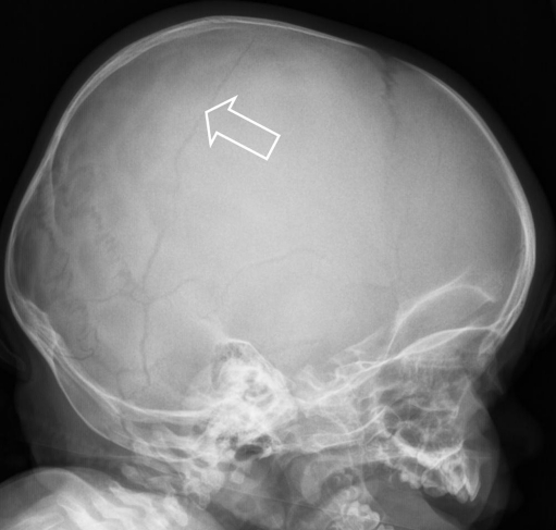
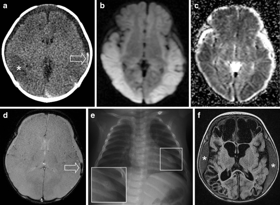
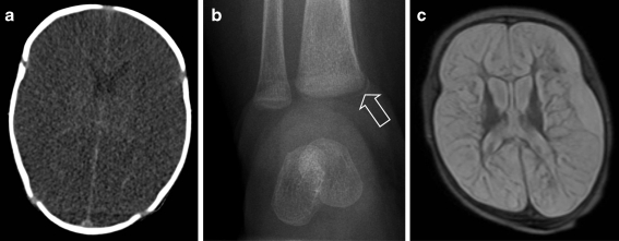
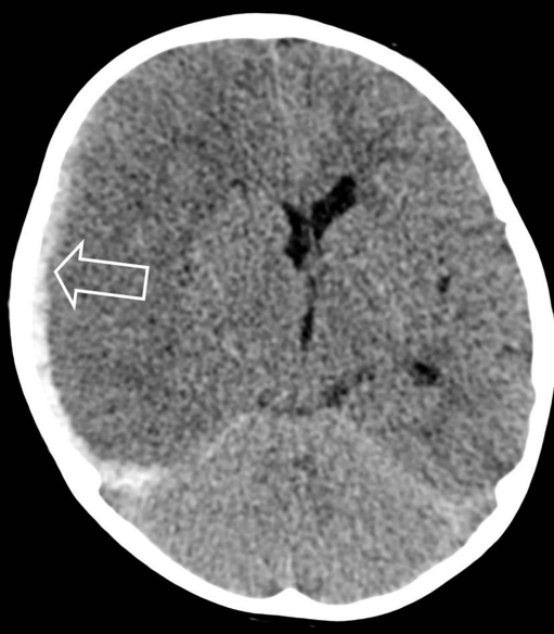
Abusive head trauma (AHT) is a relatively common cause of neurotrauma in young children. Radiology plays an important role in establishing a diagnosis and assessing a prognosis. Computed tomography (CT), followed by magnetic resonance imaging (MRI) including diffusion-weighted imaging (DWI), is the best tool for neuroimaging. There is no evidence-based approach for the follow-up of AHT; both repeat CT and MRI are currently used but literature is not conclusive. A full skeletal survey according to international guidelines should always be performed to obtain information on possible underlying bone diseases or injuries suspicious for child abuse. Cranial ultrasonography is not indicated as a diagnostic modality for the evaluation of AHT. If there is a suspicion of AHT, this should be communicated with the clinicians immediately in order to arrange protective measures as long as AHT is part of the differential diagnosis.

CONCLUSION

The final diagnosis of AHT can never be based on radiological findings only; this should always be made in a multidisciplinary team assessment where all clinical and psychosocial information is combined and judged by a group of experts in the field.

摘要

目的

探讨虐待性头部外伤(AHT)的放射学特征、诊断和治疗策略。

方法

通过对近年来发表的相关文献进行综合分析,对 AHT 的放射学表现、诊断和治疗进行了总结。

结果

AHT 是儿童神经创伤的一个常见原因。放射学在诊断和评估预后方面起着重要作用。CT 后行 MRI(包括弥散加权成像[DWI])是神经影像学的最佳工具。目前尚无 AHT 随访的循证方法;重复 CT 和 MRI 均被用于随访,但文献结论并不一致。根据国际指南,应始终进行全面的骨骼检查,以获取可能存在的潜在骨疾病或疑似虐待性损伤的信息。颅超声不适用于 AHT 的评估。如果怀疑 AHT,应立即与临床医生沟通,以便在 AHT 为鉴别诊断的一部分时安排保护措施。

结论

AHT 的最终诊断绝不能仅基于放射学发现;这应该在多学科团队评估中进行,其中所有临床和社会心理信息都结合在一起,并由该领域的一组专家进行判断。

https://cdn.ncbi.nlm.nih.gov/pmc/blobs/51de/3306566/e9b327e7fe8c/431_2011_1611_Fig6_HTML.jpg
https://cdn.ncbi.nlm.nih.gov/pmc/blobs/51de/3306566/bea0628d3aab/431_2011_1611_Fig1_HTML.jpg
https://cdn.ncbi.nlm.nih.gov/pmc/blobs/51de/3306566/f0eecd2c6a9c/431_2011_1611_Fig2_HTML.jpg
https://cdn.ncbi.nlm.nih.gov/pmc/blobs/51de/3306566/0f2db0612434/431_2011_1611_Fig3_HTML.jpg
https://cdn.ncbi.nlm.nih.gov/pmc/blobs/51de/3306566/4bc067a813be/431_2011_1611_Fig4_HTML.jpg
https://cdn.ncbi.nlm.nih.gov/pmc/blobs/51de/3306566/0916fbccd51a/431_2011_1611_Fig5_HTML.jpg
https://cdn.ncbi.nlm.nih.gov/pmc/blobs/51de/3306566/e9b327e7fe8c/431_2011_1611_Fig6_HTML.jpg

相似文献

1
Abusive head trauma Part II: radiological aspects.虐待性头部创伤 第二部分:影像学表现。
Eur J Pediatr. 2012 Apr;171(4):617-23. doi: 10.1007/s00431-011-1611-6. Epub 2011 Nov 15.
2
Unilateral hypoxic-ischemic injury in young children from abusive head trauma, lacking craniocervical vascular dissection or cord injury.幼儿因虐待性头部外伤导致的单侧缺氧缺血性损伤,无颅颈血管夹层或脊髓损伤。
Pediatr Radiol. 2008 Feb;38(2):164-74. doi: 10.1007/s00247-007-0673-0. Epub 2007 Nov 20.
3
Various Cranial and Orbital Imaging Findings in Pediatric Abusive and Non-abusive Head trauma, and Relation to Outcomes.儿童外伤性和非外伤性颅脑和眼眶影像学表现及其与结局的关系。
Clin Neuroradiol. 2019 Jun;29(2):253-261. doi: 10.1007/s00062-018-0663-7. Epub 2018 Jan 23.
4
Whole-Spine MRI in Children With Suspected Abusive Head Trauma.疑似虐待性头部创伤儿童的全脊柱 MRI。
AJR Am J Roentgenol. 2022 Jun;218(6):1074-1087. doi: 10.2214/AJR.21.26674. Epub 2022 Jan 12.
5
MRI Findings in Pediatric Abusive Head Trauma: A Review.儿童虐待性头部外伤的 MRI 表现:综述。
J Neuroimaging. 2020 Jan;30(1):15-27. doi: 10.1111/jon.12670. Epub 2019 Nov 6.
6
Serial neuroimaging in infants with abusive head trauma: timing abusive injuries.虐待性头部创伤婴儿的系列神经影像学检查:确定虐待性损伤的时间
J Neurosurg Pediatr. 2013 Aug;12(2):110-9. doi: 10.3171/2013.4.PEDS12596. Epub 2013 Jun 25.
7
Characteristics associated with spine injury on magnetic resonance imaging in children evaluated for abusive head trauma.在因虐待性头部创伤接受评估的儿童中,磁共振成像上与脊柱损伤相关的特征。
Pediatr Radiol. 2020 Jan;50(1):83-97. doi: 10.1007/s00247-019-04517-y. Epub 2020 Jan 4.
8
The neuroimaging mimics of abusive head trauma.虐待性头部创伤的神经影像学模拟。
Eur J Paediatr Neurol. 2019 Jan;23(1):19-30. doi: 10.1016/j.ejpn.2018.11.006. Epub 2018 Nov 22.
9
CNS Injuries in Abusive Head Trauma.虐待性头部创伤中的中枢神经系统损伤
AJR Am J Roentgenol. 2017 May;208(5):991-1001. doi: 10.2214/AJR.16.17602. Epub 2017 Feb 28.
10
Susceptibility-weighted imaging of retinal hemorrhages in abusive head trauma.虐待性头部创伤中视网膜出血的磁敏感加权成像
Pediatr Radiol. 2019 Feb;49(2):210-216. doi: 10.1007/s00247-018-4292-8. Epub 2018 Nov 3.

引用本文的文献

1
ESR Essentials: imaging of suspected child abuse-practice recommendations by the European Society of Paediatric Radiology.红细胞沉降率要点:欧洲儿科放射学会关于疑似虐待儿童的影像学检查实践建议
Eur Radiol. 2025 Apr;35(4):1868-1880. doi: 10.1007/s00330-024-11052-4. Epub 2024 Sep 18.
2
Abusive Head Trauma Animal Models: Focus on Biomarkers.虐待性头部创伤动物模型:关注生物标志物。
Int J Mol Sci. 2023 Feb 24;24(5):4463. doi: 10.3390/ijms24054463.
3
Factors Associated With Subdural Hygroma Following Mild Traumatic Brain Injury.轻度创伤性脑损伤后硬膜下积液的相关因素。

本文引用的文献

1
Benign external hydrocephalus: a review, with emphasis on management.良性外部性脑积水:综述,重点在于治疗。
Neurosurg Rev. 2011 Oct;34(4):417-32. doi: 10.1007/s10143-011-0327-4. Epub 2011 Jun 7.
2
Educational paper: imaging child abuse: the bare bones.教育论文:儿童虐待影像学:要点概述。
Eur J Pediatr. 2012 Feb;171(2):215-24. doi: 10.1007/s00431-011-1499-1. Epub 2011 Jun 1.
3
ACR Appropriateness Criteria(®) on suspected physical abuse-child.ACR 适宜性标准(®):疑似身体虐待儿童。
Korean J Neurotrauma. 2022 Oct 24;18(2):230-237. doi: 10.13004/kjnt.2022.18.e61. eCollection 2022 Oct.
4
Imaging of Abusive Head Trauma : A Radiologists' Perspective.虐待性头部创伤的影像学:放射科医生的视角
J Korean Neurosurg Soc. 2022 May;65(3):397-407. doi: 10.3340/jkns.2021.0297. Epub 2022 Apr 28.
5
Identification of Abusive Head Trauma in High-Risk Infants: A Cost-Effectiveness Analysis.高危婴儿虐待性头部创伤的识别:成本效益分析。
J Pediatr. 2020 Dec;227:176-183.e3. doi: 10.1016/j.jpeds.2020.06.003. Epub 2020 Jun 10.
6
Implementation of a brain injury screen MRI for infants at risk for abusive head trauma.对有虐待性头部创伤风险的婴儿实施脑损伤筛查磁共振成像。
Pediatr Radiol. 2020 Jan;50(1):75-82. doi: 10.1007/s00247-019-04506-1. Epub 2020 Jan 4.
7
Diagnostic Performance of Ultrafast Brain MRI for Evaluation of Abusive Head Trauma.用于评估虐待性头部创伤的超快脑磁共振成像的诊断性能
AJNR Am J Neuroradiol. 2017 Apr;38(4):807-813. doi: 10.3174/ajnr.A5093. Epub 2017 Feb 9.
8
Imaging of Abusive Trauma.虐待性创伤的影像学检查
Indian J Pediatr. 2016 Jun;83(6):578-88. doi: 10.1007/s12098-016-2043-0. Epub 2016 Feb 17.
9
Development of a screening MRI for infants at risk for abusive head trauma.针对有虐待性头部创伤风险的婴儿开发一种筛查性磁共振成像检查。
Pediatr Radiol. 2016 Apr;46(4):519-26. doi: 10.1007/s00247-015-3500-z. Epub 2015 Nov 20.
10
Imaging abusive head trauma: why use both computed tomography and magnetic resonance imaging?影像诊断虐待性头部外伤:为何同时使用计算机断层扫描和磁共振成像?
Pediatr Radiol. 2014 Dec;44 Suppl 4:S589-603. doi: 10.1007/s00247-014-3216-5. Epub 2014 Dec 14.
J Am Coll Radiol. 2011 Feb;8(2):87-94. doi: 10.1016/j.jacr.2010.09.008.
4
Susceptibility weighted imaging: a pictorial review.磁敏感加权成像:图文综述。
J Med Imaging Radiat Oncol. 2010 Oct;54(5):435-49. doi: 10.1111/j.1754-9485.2010.02208.x.
5
Value of cerebral microhemorrhages detected with susceptibility-weighted MR Imaging for prediction of long-term outcome in children with nonaccidental trauma.SWI 检测到的脑微出血对预测非外伤性儿童长期预后的价值。
Radiology. 2010 Sep;256(3):898-905. doi: 10.1148/radiol.10091842.
6
Abusive head trauma: judicial admissions highlight violent and repetitive shaking.虐待性头部创伤:司法认定强调暴力和反复摇晃。
Pediatrics. 2010 Sep;126(3):546-55. doi: 10.1542/peds.2009-3647. Epub 2010 Aug 9.
7
Shaken baby syndrome in Switzerland: results of a prospective follow-up study, 2002-2007.瑞士摇晃婴儿综合征:一项前瞻性随访研究的结果,2002-2007 年。
Eur J Pediatr. 2010 Aug;169(8):1023-8. doi: 10.1007/s00431-010-1175-x. Epub 2010 Mar 7.
8
Long-term skeletal findings in Menkes disease.Menkes 病的长期骨骼发现。
Pediatr Radiol. 2010 Aug;40(8):1426-9. doi: 10.1007/s00247-010-1551-8. Epub 2010 Feb 24.
9
Diffusion-weighted imaging predicts cognition in pediatric brain injury.弥散加权成像预测儿童脑损伤的认知功能。
Pediatr Neurol. 2009 Dec;41(6):406-12. doi: 10.1016/j.pediatrneurol.2009.06.002.
10
What neuroimaging should be performed in children in whom inflicted brain injury (iBI) is suspected? A systematic review.对于疑似遭受脑损伤(iBI)的儿童,应进行哪些神经影像学检查?一项系统综述。
Clin Radiol. 2009 May;64(5):473-83. doi: 10.1016/j.crad.2008.11.011. Epub 2009 Feb 20.