文献检索文档翻译深度研究
Suppr Zotero 插件Zotero 插件
邀请有礼套餐&价格历史记录

新学期,新优惠

限时优惠:9月1日-9月22日

30天高级会员仅需29元

1天体验卡首发特惠仅需5.99元

了解详情
不再提醒
插件&应用
Suppr Zotero 插件Zotero 插件浏览器插件Mac 客户端Windows 客户端微信小程序
高级版
套餐订阅购买积分包
AI 工具
文献检索文档翻译深度研究
关于我们
关于 Suppr公司介绍联系我们用户协议隐私条款
关注我们

Suppr 超能文献

核心技术专利:CN118964589B侵权必究
粤ICP备2023148730 号-1Suppr @ 2025

痛风的影像学表现——MRI、CT、双能 CT 和 US 能提供哪些信息?

Imaging in gout--what can we learn from MRI, CT, DECT and US?

机构信息

Department of Molecular Medicine and Pathology, Faculty of Medicine and Health Sciences, University of Auckland, 85 Park Road, Grafton, Auckland 1023, New Zealand.

出版信息

Arthritis Res Ther. 2011;13(6):246. doi: 10.1186/ar3489. Epub 2011 Nov 4.


DOI:10.1186/ar3489
PMID:22085684
原文链接:https://pmc.ncbi.nlm.nih.gov/articles/PMC3334625/
Abstract

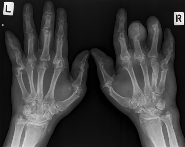
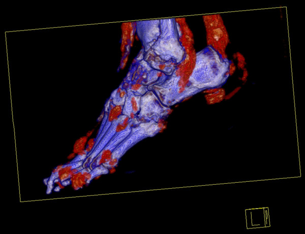
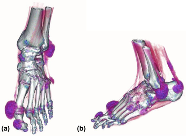
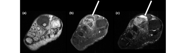
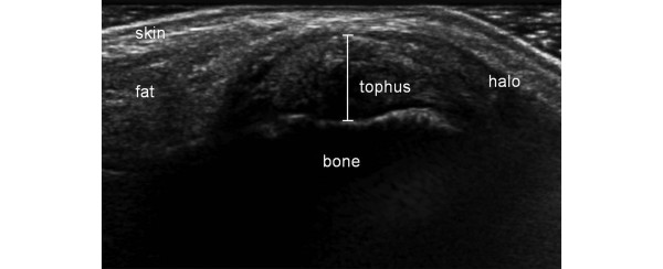
There are many exciting new applications for advanced imaging in gout. These modalities employ multiplanar imaging and allow computerized three-dimensional rendering of bone and joints (including tophi) and have the advantage of electronic data storage for later retrieval. High-resolution computed tomography has been particularly helpful in exploring the pathology of gout by investigating the relationship between bone erosions and tophi. Magnetic resonance imaging and ultrasonography can image the inflammatory nature of gouty arthropathy, revealing synovial and soft tissue inflammation, and can provide information about the composition and vascularity of tophi. Dual-energy computerized tomography is a new modality that is able to identify tophi by their chemical composition and reveal even small occult tophaceous deposits. All modalities are being investigated for their potential roles in diagnosis and could have important clinical applications in the patient for whom aspiration of monosodium urate crystals from the joint is not possible. Imaging can also provide outcome measures, such as change in tophus volume, for monitoring the response to urate-lowering therapy and this is an important application in the clinical trial setting.

摘要

在痛风的高级影像学应用方面有许多令人兴奋的新进展。这些技术采用多平面成像,并允许对骨骼和关节(包括痛风石)进行计算机化的三维渲染,具有电子数据存储的优势,便于以后检索。高分辨率计算机断层扫描(CT)通过研究骨侵蚀与痛风石之间的关系,特别有助于探索痛风的病理学。磁共振成像(MRI)和超声检查可以对痛风性关节炎的炎症性质进行成像,显示滑膜和软组织炎症,并提供关于痛风石成分和血管的信息。双能 CT 是一种新的技术,能够根据其化学成分识别痛风石,并显示甚至很小的隐匿性痛风石沉积。所有这些技术都在研究其在诊断中的潜在作用,对于那些无法从关节抽吸单钠尿酸盐晶体的患者,可能具有重要的临床应用价值。影像学还可以提供痛风石体积变化等疗效指标,用于监测降尿酸治疗的反应,这在临床试验中是一个重要的应用。

https://cdn.ncbi.nlm.nih.gov/pmc/blobs/1080/3334625/2ba1e26bce82/ar3489-5.jpg
https://cdn.ncbi.nlm.nih.gov/pmc/blobs/1080/3334625/98296a7edc9e/ar3489-1.jpg
https://cdn.ncbi.nlm.nih.gov/pmc/blobs/1080/3334625/45ff750edade/ar3489-2.jpg
https://cdn.ncbi.nlm.nih.gov/pmc/blobs/1080/3334625/3e6722bbc44c/ar3489-3.jpg
https://cdn.ncbi.nlm.nih.gov/pmc/blobs/1080/3334625/a3f40ea24c3f/ar3489-4.jpg
https://cdn.ncbi.nlm.nih.gov/pmc/blobs/1080/3334625/2ba1e26bce82/ar3489-5.jpg

相似文献

[1]
Imaging in gout--what can we learn from MRI, CT, DECT and US?

Arthritis Res Ther. 2011-11-4

[2]
New insights into an old disease: advanced imaging in the diagnosis and management of gout.

Postgrad Med J. 2012-10-30

[3]
Comparison of dual-energy CT, ultrasound and surface measurement for assessing tophus dissolution during rapid urate debulking.

Clin Rheumatol. 2017-6-16

[4]
Imaging of gout: New tools and biomarkers?

Best Pract Res Clin Rheumatol. 2016-11-12

[5]
Ultrasonography and dual-energy computed tomography provide different quantification of urate burden in gout: results from a cross-sectional study.

Arthritis Res Ther. 2017-7-21

[6]
[Multimodality imaging of chronic tophaceous gout].

Reumatismo. 2010

[7]
Dual-energy computed tomography for gout diagnosis and management.

Curr Rheumatol Rep. 2013-1

[8]
Gouty arthropathy: Review of clinico-pathologic and imaging features.

J Med Imaging Radiat Oncol. 2016-2

[9]
Imaging in gout: insights into the pathological features of disease.

Curr Opin Rheumatol. 2012-3

[10]
Relevant aspects of imaging in the diagnosis and management of gout.

Rev Bras Reumatol Engl Ed. 2017

引用本文的文献

[1]
Beyond the Signal: Imaging Insights and Diagnostic Relevance of Bone Oedema in Bone Tumours and Tumour-like Lesions.

Cancers (Basel). 2025-6-20

[2]
Patellar Non-Traumatic Pathologies: A Pictorial Review of Radiologic Findings.

Diagnostics (Basel). 2024-12-16

[3]
Spinal gout diagnosis in chiropractic practice: narrative review.

J Can Chiropr Assoc. 2023-4

[4]
Detailed analysis of the association between urate deposition and bone erosion in gout: a dual-energy computed tomography study.

Front Endocrinol (Lausanne). 2023

[5]
Unusual location of gouty arthritis with shoulder joint involvement in an elderly male patient: a rare case report.

Ann Med Surg (Lond). 2023-3-27

[6]
Surgical Treatment of Hand and Foot Gout Stone and Influence Factors on Prognosis.

Comput Math Methods Med. 2022

[7]
Multiple gouty tophi in the head and neck with normal serum uric acid: A case report and review of literatures.

World J Clin Cases. 2022-2-6

[8]
Severe erosive lesion of the glenoid in gouty shoulder arthritis: a case report and review of the literature.

BMC Musculoskelet Disord. 2021-4-12

[9]
Differentiation between infectious spondylodiscitis versus inflammatory or degenerative spinal changes: How can magnetic resonance imaging help the clinician?

Radiol Med. 2021-6

[10]
High-Resolution Peripheral Quantitative Computed Tomography for Bone Evaluation in Inflammatory Rheumatic Disease.

Front Med (Lausanne). 2020-7-15

本文引用的文献

[1]
Magnetic resonance imaging bone edema is not a major feature of gout unless there is concomitant osteomyelitis: 10-year findings from a high-prevalence population.

J Rheumatol. 2011-10-1

[2]
Methods of tophus assessment in clinical trials of chronic gout: a systematic literature review and pictorial reference guide.

Ann Rheum Dis. 2011-1-7

[3]
Ultrasound features of tophi in chronic tophaceous gout.

Skeletal Radiol. 2010-7-31

[4]
Assessment of cartilage loss at the wrist in rheumatoid arthritis using a new MRI scoring system.

Ann Rheum Dis. 2010-5-14

[5]
Dual-energy CT as a potential new diagnostic tool in the management of gout in the acute setting.

AJR Am J Roentgenol. 2010-4

[6]
Cellular characterization of the gouty tophus: a quantitative analysis.

Arthritis Rheum. 2010-5

[7]
Outcome domains for studies of acute and chronic gout.

J Rheumatol. 2009-10

[8]
An analysis of MRI and ultrasound imaging in patients with gout who have normal plain radiographs.

Rheumatology (Oxford). 2009-11

[9]
The OMERACT ultrasound task force -- Advances and priorities.

J Rheumatol. 2009-8

[10]
The OMERACT psoriatic arthritis magnetic resonance imaging scoring system (PsAMRIS): definitions of key pathologies, suggested MRI sequences, and preliminary scoring system for PsA Hands.

J Rheumatol. 2009-8

文献AI研究员

20分钟写一篇综述,助力文献阅读效率提升50倍

立即体验

用中文搜PubMed

大模型驱动的PubMed中文搜索引擎

马上搜索

推荐工具

医学文档翻译智能文献检索