Department of Biomedical Engineering, Duke University, Durham, NC 27708-0281, USA.
Ultrasound Med Biol. 2012 Jan;38(1):50-61. doi: 10.1016/j.ultrasmedbio.2011.10.002. Epub 2011 Nov 21.
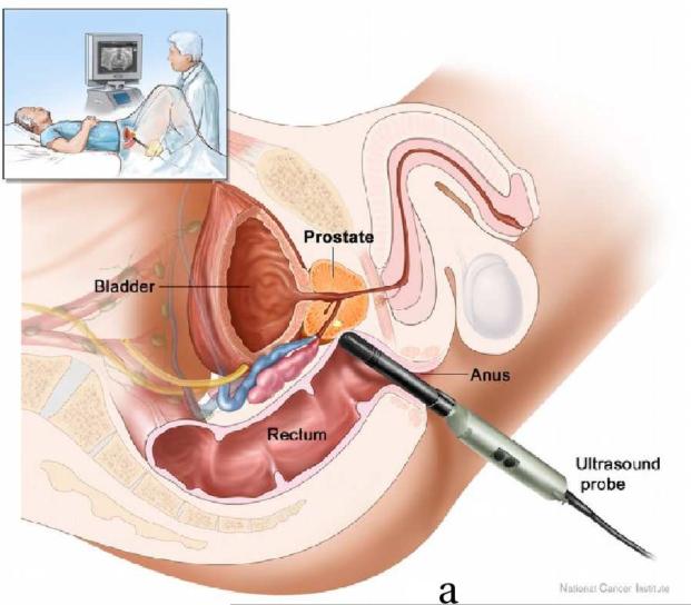
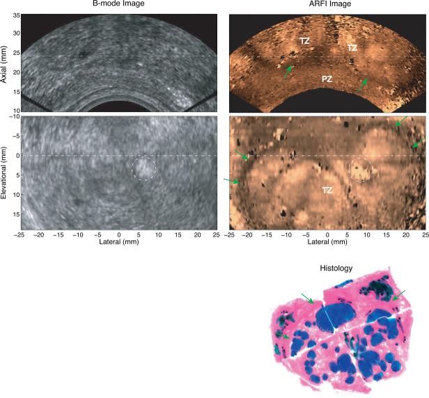
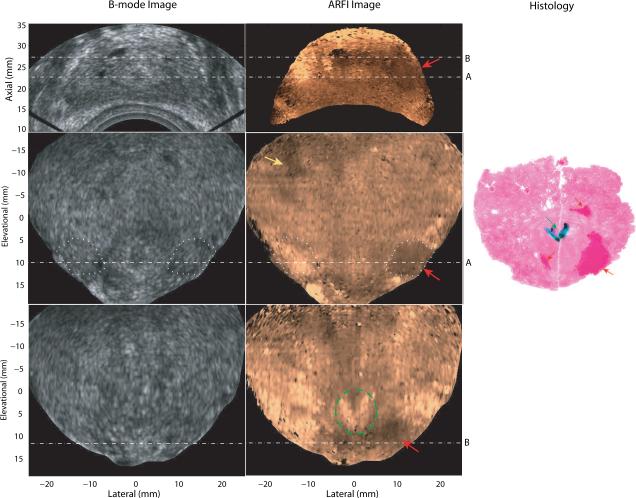
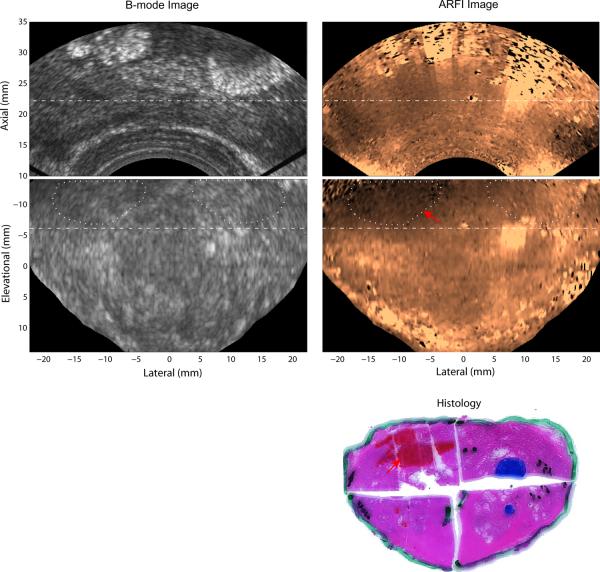
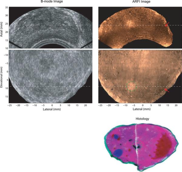
Reliably detecting prostate cancer (PCa) has been a challenge for current imaging modalities. Acoustic radiation force impulse (ARFI) imaging is an elasticity imaging method that uses remotely generated, focused acoustic beams to probe tissue stiffness. A previous study on excised human prostates demonstrated ARFI images portray various prostatic structures and has the potential to guide prostate needle biopsy with improved sampling accuracy. The goal of this study is to demonstrate the feasibility of ARFI imaging to portray internal structures and PCa in the human prostate in vivo. Custom ARFI imaging sequences were designed and implemented using a modified Siemens Antares™ scanner with a three-dimensional (3-D) wobbler, end-firing, trans-cavity transducer, EV9F4. Nineteen patients were consented and imaged immediately preceding surgical prostatectomy. Pathologies and anatomic structures were identified in histologic slides by a pathologist blinded to ARFI data and were then registered with structures found in ARFI images. The results demonstrated that when PCa is visible, it generally appears as bilaterally asymmetric stiff structures; benign prostatic hyperplasia (BPH) appears heterogeneous with a nodular texture; the verumontanum and ejaculatory ducts appears softer compared with surrounding tissue, which form a unique 'V' shape; and the boundary of the transitional zone (TZ) forms a stiff rim separating the TZ from the peripheral zone (PZ). These characteristic appearances of prostatic structures are consistent with those found in our previous study of prostate ARFI imaging on excised human prostates. Compared with the matched B-mode images, ARFI images, in general, portray prostate structures with higher contrast. With the end-firing transducer used for this study, ARFI depth penetration was limited to 22 mm. Image contrast and resolution were decreased as compared with the previous ex vivo study due to the small transducer aperture. Even with these limitations, this study suggests ARFI imaging holds promise for guidance of targeted prostate needle biopsy and focal therapy, as well as aiding assessment of changes during watchful waiting/active surveillance.
准确检测前列腺癌 (PCa) 一直是当前成像方式的挑战。声辐射力脉冲 (ARFI) 成像是一种弹性成像方法,它使用远程产生的聚焦声束来探测组织的硬度。之前对切除的人类前列腺的研究表明,ARFI 图像可以描绘各种前列腺结构,并有可能通过提高采样准确性来指导前列腺针活检。本研究旨在证明 ARFI 成像在体内描绘人类前列腺内部结构和 PCa 的可行性。使用带有三维 (3-D) 摆动器、端射、腔内换能器 EV9F4 的改良 Siemens Antares™ 扫描仪设计并实现了定制的 ARFI 成像序列。19 名患者在接受前列腺切除术之前同意并进行了成像。病理学家在组织学切片中识别出病理和解剖结构,然后将这些结构与 ARFI 图像中发现的结构进行注册。结果表明,当 PCa 可见时,它通常表现为双侧不对称的硬结构;良性前列腺增生 (BPH) 表现为不均匀的结节状质地;与周围组织相比,精阜和射精管显得更软,形成独特的“V”形;而移行带 (TZ) 的边界形成一个坚硬的边缘,将 TZ 与周围带 (PZ) 分开。这些前列腺结构的特征外观与我们之前对切除的人类前列腺 ARFI 成像的研究一致。与匹配的 B 模式图像相比,ARFI 图像通常可以更清晰地描绘前列腺结构。与之前的离体研究相比,由于换能器孔径较小,该研究中使用的端射换能器限制了 ARFI 的深度穿透,仅为 22mm。由于换能器孔径较小,图像对比度和分辨率均低于之前的离体研究。尽管存在这些限制,但本研究表明 ARFI 成像有望指导靶向前列腺针活检和聚焦治疗,并有助于评估等待观察/主动监测期间的变化。