Spitzer Philipp, Schieb Heinke, Kamrowski-Kruck Heike, Otto Markus, Chiasserini Davide, Parnetti Lucilla, Herukka Sanna-Kaisa, Schuchhardt Johannes, Wiltfang Jens, Klafki Hans-Wolfgang
Department of Psychiatry and Psychotherapy, Laboratory for Molecular Neurobiology, LVR-Klinikum Essen, University of Duisburg-Essen, Virchowstraße 174, 45147 Essen, Germany.
Int J Alzheimers Dis. 2011;2011:739847. doi: 10.4061/2011/739847. Epub 2011 Nov 24.
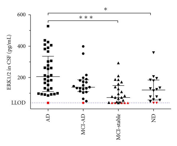
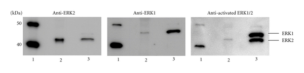
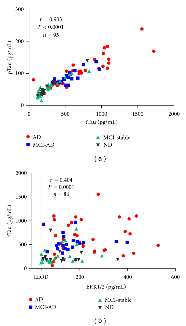
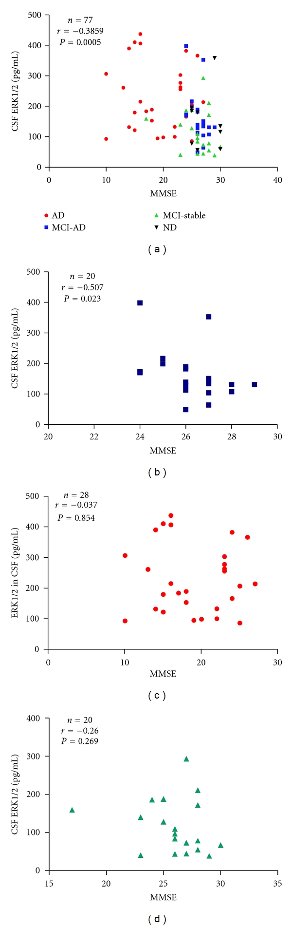
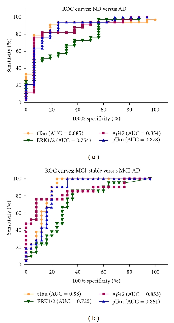
Cerebrospinal fluid (CSF) samples from 33 patients with Alzheimer dementia (AD), 21 patients with mild cognitive impairment who converted to AD during followup (MCI-AD), 25 patients with stable mild cognitive impairment (MCI-stable), and 16 nondemented subjects (ND) were analyzed with a chemiluminescence immunoassay to assess the levels of the mitogen-activated protein kinase ERK1/2 (extracellular signal-regulated kinase 1/2). The results were evaluated in relation to total Tau (tTau), phosphorylated Tau (pTau), and beta-amyloid 42 peptide (Aβ42). CSF-ERK1/2 was significantly increased in the AD group as compared to stable MCI patients and the ND group. Western blot analysis of a pooled cerebrospinal fluid sample revealed that both isoforms, ERK1 and ERK2, and low amounts of doubly phosphorylated ERK2 were detectable. As a predictive diagnostic AD biomarker, CSF-ERK1/2 was inferior to tTau, pTau, and Aβ42.
采用化学发光免疫分析法对33例阿尔茨海默病(AD)患者、21例随访期间转化为AD的轻度认知障碍患者(MCI-AD)、25例稳定的轻度认知障碍患者(MCI-稳定)和16例非痴呆受试者(ND)的脑脊液(CSF)样本进行分析,以评估丝裂原活化蛋白激酶ERK1/2(细胞外信号调节激酶1/2)的水平。结合总 Tau(tTau)、磷酸化 Tau(pTau)和β淀粉样蛋白42肽(Aβ42)对结果进行评估。与稳定的MCI患者和ND组相比,AD组的脑脊液ERK1/2显著升高。对一份合并的脑脊液样本进行蛋白质印迹分析显示,可检测到ERK1和ERK2这两种亚型以及少量双磷酸化的ERK2。作为预测性诊断AD生物标志物,脑脊液ERK1/2不如tTau、pTau 和 Aβ42。