Center for Neural Science, New York University, New York, NY, USA.
J Neurophysiol. 2012 Mar;107(5):1275-90. doi: 10.1152/jn.00867.2011. Epub 2011 Dec 7.
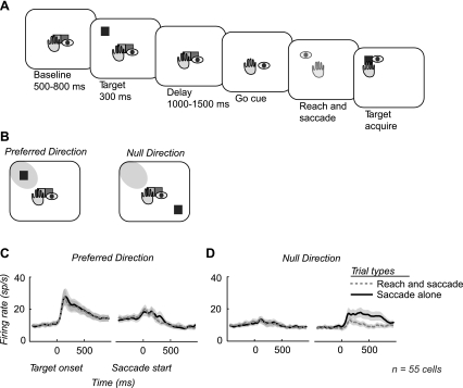
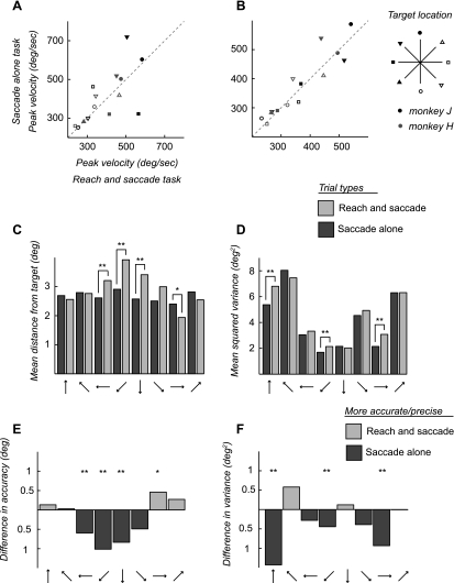
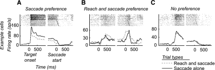
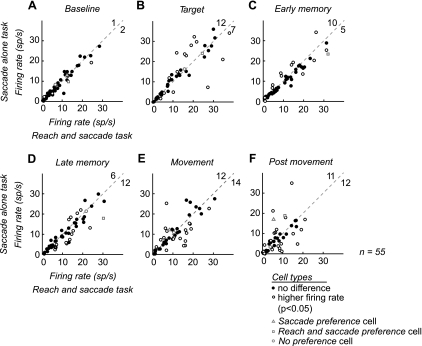
The posterior parietal cortex is situated between visual and motor areas and supports coordinated visually guided behavior. Area LIP in the intraparietal sulcus contains representations of visual space and has been extensively studied in the context of saccades. However, area LIP has not been studied during coordinated movements, so it is not known whether saccadic representations in area LIP are influenced by coordinated behavior. Here, we studied spiking and local field potential (LFP) activity in area LIP while subjects performed coordinated reaches and saccades or saccades alone to remembered target locations to test whether activity in area LIP is influenced by the presence of a coordinated reach. We find that coordination significantly changes the activity of individual neurons in area LIP, increasing or decreasing the firing rate when a reach is made with a saccade compared with when a saccade is made alone. Analyzing spike-field coherence demonstrates that area LIP neurons whose firing rate is suppressed during the coordinated task have activity temporally correlated with nearby LFP activity, which reflects the synaptic activity of populations of neurons. Area LIP neurons whose firing rate increases during the coordinated task do not show significant spike-field coherence. Furthermore, LFP power in area LIP is suppressed and does not increase when a coordinated reach is made with a saccade. These results demonstrate that area LIP neurons display different responses to coordinated reach and saccade movements, and that different spike rate responses are associated with different patterns of correlated activity. The population of neurons whose firing rate is suppressed is coherently active with local populations of LIP neurons. Overall, these results suggest that area LIP plays a role in coordinating visually guided actions through suppression of coherent patterns of saccade-related activity.
顶叶后皮质位于视觉和运动区域之间,支持协调的视觉引导行为。顶内沟中的 LIP 区域包含视觉空间的表示,并且在扫视的背景下已经得到了广泛的研究。然而,在协调运动期间尚未研究 LIP 区域,因此尚不清楚 LIP 区域中的扫视表示是否受到协调行为的影响。在这里,我们研究了主体进行协调的伸展和扫视或单独扫视到记忆目标位置时 LIP 区域中的尖峰和局部场电位(LFP)活动,以测试 LIP 区域中的活动是否受到协调伸展的影响。我们发现,协调运动显著改变了 LIP 区域中单个神经元的活动,与单独进行扫视相比,在进行带有扫视的伸展时,神经元的发射率增加或减少。分析尖峰场相干性表明,在协调任务期间,其发射率被抑制的 LIP 区域神经元的活动与附近的 LFP 活动具有时间相关性,这反映了神经元群体的突触活动。在协调任务期间,其发射率增加的 LIP 区域神经元没有显示出明显的尖峰场相干性。此外,当进行带有扫视的协调伸展时,LIP 区域中的 LFP 功率被抑制且不会增加。这些结果表明,LIP 区域神经元对协调的伸展和扫视运动表现出不同的反应,并且不同的尖峰率反应与不同的相关活动模式相关。发射率受到抑制的神经元群体与 LIP 神经元的局部群体具有相干活性。总体而言,这些结果表明,LIP 区域通过抑制与扫视相关的活动的相干模式,在协调视觉引导的动作中发挥作用。