Department of Medical Environmental Biology, Chung-Ang University College of Medicine, Seoul, Republic of Korea.
PLoS Negl Trop Dis. 2011 Dec;5(12):e1414. doi: 10.1371/journal.pntd.0001414. Epub 2011 Dec 13.
Adult Clonorchis sinensis live in the bile duct and cause clonorchiasis. It is known that the C. sinensis metacercariae excyst in the duodenum and migrate up to the bile duct through the common bile duct. However, no direct evidence is available on the in vivo migration of newly excysted C. sinensis juveniles (CsNEJs). Advanced imaging technologies now allow the in vivo migration and localization to be visualized. In the present study, we sought to determine how sensitively CsNEJs respond to bile and how fast they migrate to the intrahepatic bile duct using PET-CT.
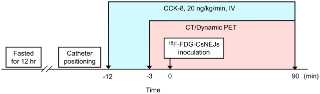
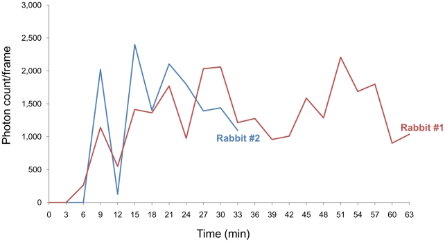
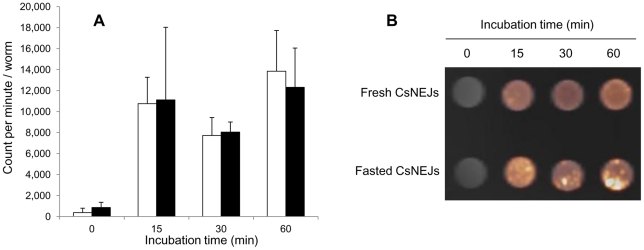
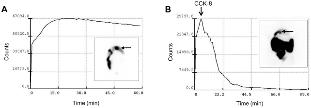
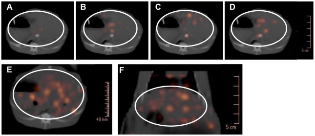
METHODOLOGY/PRINCIPAL FINDINGS: CsNEJs were radiolabeled with (18)F-fluorodeoxyglucose ((18)F-FDG). Rabbits with a gallbladder contraction response to cholecystokinin-8 (CCK-8) injection were pre-screened using cholescintigraphy. In these rabbits, gallbladders contracted by 50% in volume at an average of 11.5 min post-injection. The four rabbits examined were kept anesthetized and a catheter inserted into the mid duodenum. Gallbladder contraction was stimulated by injecting CCK-8 (20 ng/kg every minute) over the experiment. Anatomical images were acquired by CT initially and dynamic PET was then carried out for 90 min with a 3-min acquisition per frame. Twelve minutes after CCK-8 injection, about 3,000 (18)F-FDG-labeled CsNEJs were inoculated into the mid duodenum through the catheter. Photon signals were detected in the liver 7-9 min after CsNEJs inoculation, and these then increased in the whole liver with stronger intensity in the central area, presenting that the CsNEJs were arriving at the intrahepatic bile ducts.
In the duodenum, CsNEJs immediately sense bile and migrate quickly with bile-chemotaxis to reach the intrahepatic bile ducts by way of the ampulla of Vater.
成体华支睾吸虫生活在胆管中,引起华支睾吸虫病。已知华支睾吸虫囊蚴在十二指肠中脱囊,并通过胆总管迁移至胆管。然而,尚无直接证据表明新脱囊的华支睾吸虫幼虫(CsNEJs)的体内迁移。先进的成像技术现在可以使体内迁移和定位可视化。在本研究中,我们试图确定 CsNEJs 对胆汁的敏感性以及它们通过 PET-CT 快速迁移到肝内胆管的速度。
方法/主要发现:CsNEJs 用(18)F-氟脱氧葡萄糖((18)F-FDG)进行放射性标记。使用肝胆闪烁显像术对胆囊对胆囊收缩素-8(CCK-8)注射有反应的兔子进行预筛选。在这些兔子中,胆囊在注射后平均 11.5 分钟时体积收缩了 50%。检查的四只兔子保持麻醉状态,并将导管插入中十二指肠。通过注射 CCK-8(每分钟 20ng/kg)刺激胆囊收缩,进行实验。最初通过 CT 采集解剖图像,然后进行 90 分钟的动态 PET,每 3 分钟采集一帧。在 CCK-8 注射后 12 分钟,通过导管将约 3000 个(18)F-FDG 标记的 CsNEJs 接种到中十二指肠。CsNEJs 接种后 7-9 分钟在肝脏中检测到光子信号,然后在整个肝脏中信号增加,强度在中央区域更强,表明 CsNEJs 到达肝内胆管。
在十二指肠中,CsNEJs 立即感知胆汁,并通过胆汁趋化性快速迁移,通过壶腹到达肝内胆管。