Department of Cellular and Molecular Medicine, Carol Davila University of Medicine and Pharmacy, Bucharest, Romania.
J Cell Mol Med. 2012 Apr;16(4):701-7. doi: 10.1111/j.1582-4934.2011.01505.x.
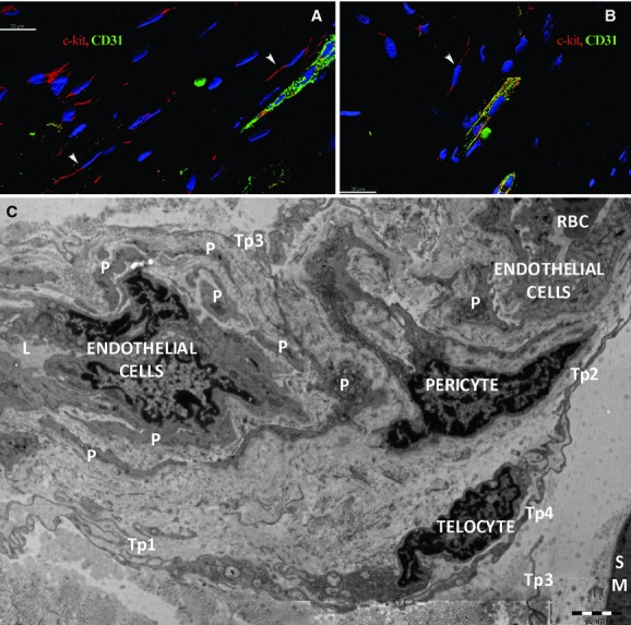
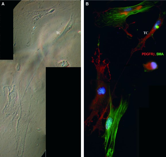
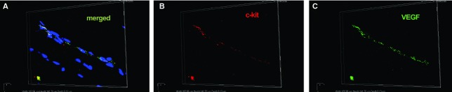
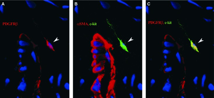
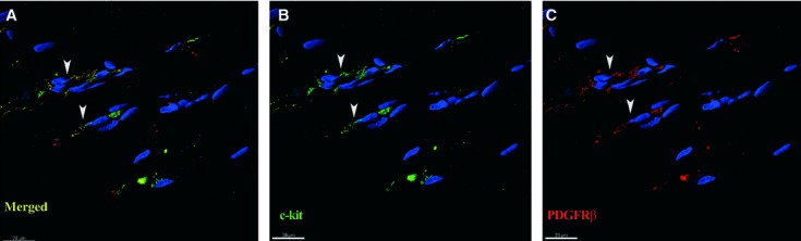
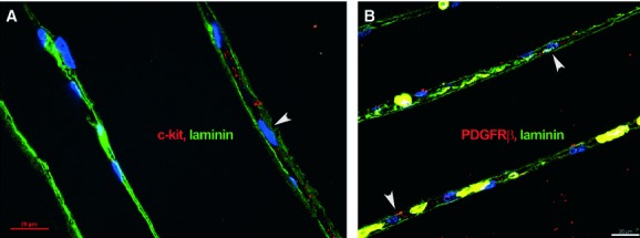
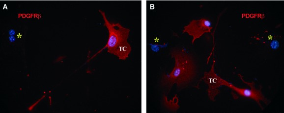
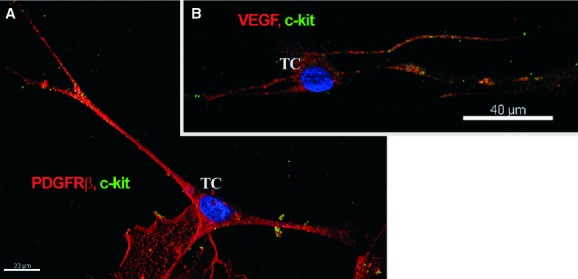
Telocytes (TCs) represent a new cell type recently described in mammalian skeletal muscle interstitium as well as in other organs. These have a specific morphology and phenotype, both in situ and in vitro. Telocytes are cells with long and slender cell prolongations, in contact with other interstitial cells, nerve fibres, blood capillaries and resident stem cells in niches. Our aim was to investigate the potential contribution of TCs to micro-vascular networks by immunofluorescent labelling of specific angiogenic growth factors and receptors. We found that in human skeletal muscle TCs were constantly located around intermediate and small blood vessels and endomysial capillaries. Epi-fluorescence and laser confocal microscopy showed that TCs express c-kit, platelet-derived growth factor receptor (PDGFR)-β and VEGF, both in situ and in vitro. Telocytes were constantly located in the perivascular or pericapillary space, as confirmed by double staining of c-kit/CD31, PDGFR-β/CD31 and PDGFR-β/α-smooth muscle actin, respectively. Electron microscopy (EM) differentiated between pericytes and other cell types. Laminin labelling showed that TCs are not enclosed or surrounded by a basal lamina in contrast to mural cells. In conclusion, a) PDGFR-β could be used as a marker for TCs and b) TCs are presumably a transitional population in the complex process of mural cell recruitment during angiogenesis and vascular remodelling.
间质细胞(TCs)是一种新型细胞,最近在哺乳动物骨骼肌间质以及其他器官中被描述。这些细胞具有特定的形态和表型,无论是在原位还是在体外。TCs 是具有长而细的细胞突起的细胞,与其他间质细胞、神经纤维、毛细血管和龛位中的常驻干细胞接触。我们的目的是通过免疫荧光标记特定的血管生成生长因子和受体来研究 TCs 对微血管网络的潜在贡献。我们发现,在人类骨骼肌中,TCs 始终位于中间和小血管以及肌内膜毛细血管周围。荧光和激光共聚焦显微镜显示,TCs 在原位和体外均表达 c-kit、血小板衍生生长因子受体(PDGFR)-β 和 VEGF。TCs 始终位于血管周围或血管周围腔中,这通过 c-kit/CD31、PDGFR-β/CD31 和 PDGFR-β/α-平滑肌肌动蛋白的双重染色得到证实。电子显微镜(EM)区分了周细胞和其他细胞类型。层粘连蛋白标记表明,与壁细胞不同,TCs 没有被基底膜包围或包裹。总之,a)PDGFR-β 可作为 TCs 的标志物,b)TCs 可能是血管生成和血管重塑过程中募集壁细胞的复杂过程中的过渡群体。