• 文献检索
  • 文档翻译
  • 深度研究
  • 学术资讯
  • Suppr Zotero 插件Zotero 插件
  • 邀请有礼
  • 套餐&价格
  • 历史记录
应用&插件
Suppr Zotero 插件Zotero 插件浏览器插件Mac 客户端Windows 客户端微信小程序
定价
高级版会员购买积分包购买API积分包
服务
文献检索文档翻译深度研究API 文档MCP 服务
关于我们
关于 Suppr公司介绍联系我们用户协议隐私条款
关注我们

Suppr 超能文献

核心技术专利:CN118964589B侵权必究
粤ICP备2023148730 号-1Suppr @ 2026

文献检索

告别复杂PubMed语法,用中文像聊天一样搜索,搜遍4000万医学文献。AI智能推荐,让科研检索更轻松。

立即免费搜索

文件翻译

保留排版,准确专业,支持PDF/Word/PPT等文件格式,支持 12+语言互译。

免费翻译文档

深度研究

AI帮你快速写综述,25分钟生成高质量综述,智能提取关键信息,辅助科研写作。

立即免费体验

采用 8 点“渐进测试”测量原发性腕管综合征的超声正中神经横截面积:神经传导研究严重程度与临床症状持续时间的相关性。

Ultrasonographic median nerve cross-section areas measured by 8-point "inching test" for idiopathic carpal tunnel syndrome: a correlation of nerve conduction study severity and duration of clinical symptoms.

机构信息

Department of Neurology, Kaohsiung Chang Gung Memorial Hospital and Chang Gung University College of Medicine, Kaohsiung, Taiwan.

出版信息

BMC Med Imaging. 2011 Dec 21;11:22. doi: 10.1186/1471-2342-11-22.

DOI:10.1186/1471-2342-11-22
PMID:22189264
原文链接:https://pmc.ncbi.nlm.nih.gov/articles/PMC3292452/
Abstract

BACKGROUND

Incremental palmar stimulation of the median nerve sensory conduction at the wrist, the "inching test", provides an assessment with reference to segments proximal and distal to the entrapment. This study used high-resolution ultrasonography (US) to measure the median nerve's cross-section areas (CSAs) like the "inching test" and to correlate with the nerve conduction study (NCS) severity and duration of carpal tunnel syndrome (CTS).

METHODS

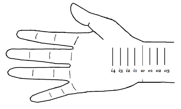
Two hundred and twelve (212) "CTS-hands" from 135 CTS patients and 50 asymptomatic hands ("A-hands") from 25 control individuals were enrolled. The median nerve CSAs were measured at the 8-point marked as i4, i3, i2, i1, w, o1, o2, and 03 in inching test. The NCS severities were classified into six groups based on motor and sensory responses (i.e., negative, minimal, mild, moderate, severe, and extreme). Results of US studies were compared in terms of NCS severity and duration of clinical CTS symptoms.

RESULTS

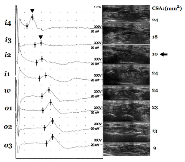
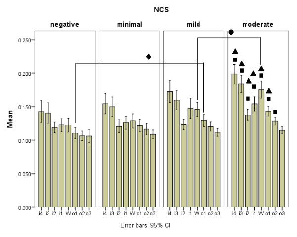
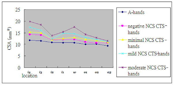
There was significantly larger CSA of the NCS negative group of "CTS-hands" than of "A-hands". The cut-off values of the CSAs of the NCS negative CTS group were 12.5 mm2, 11.5 mm2 and 10.1 mm2 at the inlet, wrist crease, and outlet, respectively. Of the 212 "CTS-hands", 32 were NCS negative while 40 had minimal, 43 mild, 85 moderate, 10 severe, and two extreme NCS severities. The CSAs of "CTS-hands" positively correlated with different NCS severities and with the duration of CTS symptoms. By duration of clinical symptoms, 12 of the 212 "CTS-hands" were in the 1 month group; 82 in >1 month and ≤ 12 months group, and 118 in >12 months group. In "inching test", segments i4-i3 and i3-i2 were the most common "positive-site". The corresponding CSAs measured at i4 and i3, but not at i2, were significantly larger than those measured at points that were not "positive-site".

CONCLUSIONS

Using the 8-point measurement of the median nerve CSA from inlet to outlet similar to the "inching test" has positive correlations with NCS severity and duration of CTS clinical symptoms, and can provide more information on anatomic changes. Combined NCS and US studies using the 8-point measurement may have a higher positive rate than NCS alone for diagnosing CTS.

摘要

背景

在腕部对正中神经感觉传导进行渐进性手掌刺激,即“逐渐递增测试”,可以提供一种与卡压部位近端和远端相关的评估。本研究使用高分辨率超声(US)测量正中神经的横截面积(CSA),类似于“逐渐递增测试”,并将其与神经传导研究(NCS)严重程度和腕管综合征(CTS)的持续时间相关联。

方法

共纳入 135 例 CTS 患者的 212 只“CTS 手”和 25 名对照个体的 50 只无症状手(“A 手”)。在逐渐递增测试中,在 i4、i3、i2、i1、w、o1、o2 和 03 8 个标记点测量正中神经 CSA。根据运动和感觉反应,将 NCS 严重程度分为 6 组(即阴性、轻微、轻度、中度、重度和极度)。比较 US 研究结果在 NCS 严重程度和 CTS 临床症状持续时间方面的差异。

结果

“CTS 手”中 NCS 阴性组的 CSA 明显大于“A 手”。NCS 阴性 CTS 组 CSA 的截断值分别为 12.5mm2、11.5mm2 和 10.1mm2,位于入口处、腕部褶皱和出口处。在 212 只“CTS 手”中,32 只 NCS 阴性,40 只 NCS 轻微,43 只 NCS 轻度,85 只 NCS 中度,10 只 NCS 重度,2 只 NCS 极度。“CTS 手”的 CSA 与不同的 NCS 严重程度和 CTS 症状持续时间呈正相关。根据临床症状持续时间,212 只“CTS 手”中,12 只为 1 个月组;82 只为>1 个月且≤12 个月组,118 只为>12 个月组。在“逐渐递增测试”中,i4-i3 和 i3-i2 段是最常见的“阳性部位”。在 i4 和 i3 处测量到的 CSA 明显大于在 i2 处测量到的 CSA,而在 i2 处测量到的 CSA 明显大于不在“阳性部位”处测量到的 CSA。

结论

使用类似于“逐渐递增测试”的从中部到出口的正中神经 CSA 的 8 点测量与 NCS 严重程度和 CTS 临床症状持续时间呈正相关,并能提供有关解剖结构变化的更多信息。与单独使用 NCS 相比,使用 8 点测量进行 NCS 和 US 联合研究可能具有更高的阳性率,用于诊断 CTS。

https://cdn.ncbi.nlm.nih.gov/pmc/blobs/10af/3292452/3862341dad8e/1471-2342-11-22-4.jpg
https://cdn.ncbi.nlm.nih.gov/pmc/blobs/10af/3292452/85bf68c5bf1c/1471-2342-11-22-1.jpg
https://cdn.ncbi.nlm.nih.gov/pmc/blobs/10af/3292452/9f954a7c7e7b/1471-2342-11-22-2.jpg
https://cdn.ncbi.nlm.nih.gov/pmc/blobs/10af/3292452/25ddcf142da8/1471-2342-11-22-3.jpg
https://cdn.ncbi.nlm.nih.gov/pmc/blobs/10af/3292452/3862341dad8e/1471-2342-11-22-4.jpg

相似文献

1
Ultrasonographic median nerve cross-section areas measured by 8-point "inching test" for idiopathic carpal tunnel syndrome: a correlation of nerve conduction study severity and duration of clinical symptoms.采用 8 点“渐进测试”测量原发性腕管综合征的超声正中神经横截面积:神经传导研究严重程度与临床症状持续时间的相关性。
BMC Med Imaging. 2011 Dec 21;11:22. doi: 10.1186/1471-2342-11-22.
2
Ultrasonographic assessment of carpal tunnel syndrome of mild and moderate severity in diabetic patients by using an 8-point measurement of median nerve cross-sectional areas.采用 8 点测量正中神经横截面积的方法对糖尿病患者轻、中度腕管综合征进行超声评估。
BMC Med Imaging. 2012 Jul 7;12:15. doi: 10.1186/1471-2342-12-15.
3
Diagnosis of carpal tunnel syndrome assessed using high-frequency ultrasonography: cross-section areas of 8-site median nerve.使用高频超声评估腕管综合征:8个部位正中神经的横截面积
Clin Rheumatol. 2016 Oct;35(10):2557-64. doi: 10.1007/s10067-016-3214-0. Epub 2016 Feb 22.
4
Severity of Carpal tunnel syndrome assessed with high frequency ultrasonography.高频超声评估腕管综合征的严重程度。
Rheumatol Int. 2010 Apr;30(6):761-5. doi: 10.1007/s00296-009-1061-x. Epub 2009 Jul 11.
5
The diagnostic value of ultrasound compared with nerve conduction velocity in carpal tunnel syndrome.超声与神经传导速度在腕管综合征中的诊断价值比较。
Int J Rheum Dis. 2014 Jul;17(6):612-20. doi: 10.1111/1756-185X.12310. Epub 2014 Feb 20.
6
Physiological Localization by Sensory and Motor Inching Studies and Structural Abnormalities Detected by Ultrasonographic Changes in Carpal Tunnel Syndrome.通过感觉和运动渐进性研究进行的生理定位以及腕管综合征超声变化检测到的结构异常
Arch Phys Med Rehabil. 2022 Mar;103(3):494-504.e1. doi: 10.1016/j.apmr.2021.08.001. Epub 2021 Aug 12.
7
The significance of ultrasonographic carpal tunnel outlet measurements in the diagnosis of carpal tunnel syndrome.超声测量腕管出口在腕管综合征诊断中的意义。
Clin Neurophysiol. 2016 Dec;127(12):3516-3523. doi: 10.1016/j.clinph.2016.09.015. Epub 2016 Oct 3.
8
Sonographic cross-sectional area measurement in carpal tunnel syndrome patients: can delta and ratio calculations predict severity compared to nerve conduction studies?腕管综合征患者的超声横截面积测量:与神经传导研究相比,差值和比值计算能否预测病情严重程度?
Eur Radiol. 2015 Aug;25(8):2419-27. doi: 10.1007/s00330-015-3649-8. Epub 2015 Apr 10.
9
Does measuring the median nerve at the carpal tunnel outlet improve ultrasound CTS diagnosis?在腕管出口处测量正中神经是否能改善超声对腕管综合征的诊断?
J Neurol Sci. 2014 Apr 15;339(1-2):47-51. doi: 10.1016/j.jns.2014.01.018. Epub 2014 Jan 17.
10
Sonographic measurement of cross-sectional area of the median nerve in the diagnosis of carpal tunnel syndrome: correlation with nerve conduction studies.超声测量正中神经横截面积在腕管综合征诊断中的应用:与神经传导研究的相关性
J Clin Ultrasound. 2009 Mar-Apr;37(3):125-31. doi: 10.1002/jcu.20551.

引用本文的文献

1
Long-term patient-centered outcomes following carpal tunnel release surgery: a 10-year follow-up.腕管松解术后以患者为中心的长期疗效:一项10年随访研究
Langenbecks Arch Surg. 2025 Apr 15;410(1):126. doi: 10.1007/s00423-025-03664-1.
2
Diagnostic Value of Measurements of Median Nerve Diameter at the Site of the Maximal Stenosis in Carpal Tunnel Syndrome.腕管综合征最大狭窄部位正中神经直径测量的诊断价值
Int J Gen Med. 2025 Jan 23;18:357-369. doi: 10.2147/IJGM.S502961. eCollection 2025.
3
Exploring ultrasound and electromyography for carpal tunnel syndrome diagnosis: a comprehensive comparative study and implications for occupational medicine.

本文引用的文献

1
The ultrasonographic correlates of carpal tunnel syndrome in patients with normal electrodiagnostic tests.正常电诊断检查患者的腕管综合征的超声相关性。
Radiol Med. 2011 Apr;116(3):489-96. doi: 10.1007/s11547-011-0632-6. Epub 2011 Feb 1.
2
Median nerve conduction study through the carpal tunnel using segmental nerve length measured by ultrasonographic and conventional tape methods.腕管内正中神经传导研究采用超声和常规带测量的节段神经长度。
Arch Phys Med Rehabil. 2011 Jan;92(1):1-6. doi: 10.1016/j.apmr.2010.09.012.
3
Diagnostic value of cross-sectional area of median nerve in grading severity of carpal tunnel syndrome.
探索超声和肌电图在腕管综合征诊断中的应用:一项全面的比较研究及其对职业医学的启示
Front Neurol. 2024 Dec 11;15:1490873. doi: 10.3389/fneur.2024.1490873. eCollection 2024.
4
Nerve ultrasonographic findings in diabetes mellitus are determined by anatomical location and type of diabetes.糖尿病患者的神经超声检查结果取决于解剖位置和糖尿病类型。
Clin Neurophysiol Pract. 2023 May 24;8:115-122. doi: 10.1016/j.cnp.2023.04.004. eCollection 2023.
5
Sonographic reference values for median nerve cross-sectional area: A meta-analysis of data from healthy individuals.正中神经横截面积的超声参考值:对健康个体数据的荟萃分析。
J Diagn Med Sonogr. 2023 Sep;39(5):492-506. doi: 10.1177/87564793231176009. Epub 2023 Jun 6.
6
Ratio and difference of the cross-sectional area of median nerve to ulnar nerve in diagnosing carpal tunnel syndrome: a case control study.正中神经与尺神经横截面积比值在腕管综合征诊断中的作用:一项病例对照研究。
BMC Med Imaging. 2019 Jul 4;19(1):52. doi: 10.1186/s12880-019-0351-3.
7
Sonographic assessment of carpal tunnel syndrome in diabetic patients with and without polyneuropathy.超声评估合并或不合并多发性神经病的糖尿病患者的腕管综合征
Medicine (Baltimore). 2018 Jun;97(24):e11104. doi: 10.1097/MD.0000000000011104.
8
Anatomical variations of the pronator teres muscle in a Central European population and its clinical significance.中欧人群中旋前圆肌的解剖变异及其临床意义。
Anat Sci Int. 2018 Mar;93(2):299-306. doi: 10.1007/s12565-017-0413-y. Epub 2017 Aug 28.
9
Ultrasound elastography for carpal tunnel pressure measurement: A cadaveric validation study.用于腕管压力测量的超声弹性成像:一项尸体验证研究。
J Orthop Res. 2018 Jan;36(1):477-483. doi: 10.1002/jor.23658. Epub 2017 Aug 14.
10
The Prevalence of Pronator Teres among Patients with Carpal Tunnel Syndrome: Cross-sectional Study.腕管综合征患者中旋前圆肌的患病率:横断面研究。
Int J Biomed Sci. 2016 Sep;12(3):89-94.
正中神经截面积在腕管综合征严重程度分级中的诊断价值。
Arch Iran Med. 2010 Nov;13(6):516-21.
4
Repeatability of ultrasonographic median nerve measures.超声测量正中神经重复性研究。
Muscle Nerve. 2010 Jun;41(6):767-73. doi: 10.1002/mus.21619.
5
Usefulness of additional measurements of the median nerve with ultrasonography.超声检查正中神经附加测量的有用性。
Neurol Sci. 2010 Dec;31(6):721-5. doi: 10.1007/s10072-010-0258-9. Epub 2010 Apr 29.
6
Severity of Carpal tunnel syndrome assessed with high frequency ultrasonography.高频超声评估腕管综合征的严重程度。
Rheumatol Int. 2010 Apr;30(6):761-5. doi: 10.1007/s00296-009-1061-x. Epub 2009 Jul 11.
7
Carpal tunnel syndrome: ultrasound, neurophysiology, clinical and patient-oriented assessment.腕管综合征:超声、神经生理学、临床及以患者为导向的评估
Clin Neurophysiol. 2008 Sep;119(9):2064-9. doi: 10.1016/j.clinph.2008.05.004. Epub 2008 Jul 11.
8
The ultrasonographic wrist-to-forearm median nerve area ratio in carpal tunnel syndrome.腕管综合征中超声检查的腕部至前臂正中神经面积比
Clin Neurophysiol. 2008 Jun;119(6):1353-7. doi: 10.1016/j.clinph.2008.01.101. Epub 2008 Apr 1.
9
Diagnostic utility of ultrasonography versus nerve conduction studies in mild carpal tunnel syndrome.超声检查与神经传导研究在轻度腕管综合征中的诊断效用
Arthritis Rheum. 2008 Mar 15;59(3):357-66. doi: 10.1002/art.23317.
10
High-resolution sonography versus EMG in the diagnosis of carpal tunnel syndrome.高分辨率超声检查与肌电图在腕管综合征诊断中的比较
J Neurol Neurosurg Psychiatry. 2008 Jan;79(1):63-7. doi: 10.1136/jnnp.2007.115337. Epub 2007 Apr 30.