Department of Radiology, University Medical Center Utrecht, Heidelberglaan 100, E01.132, PO BOX 85500, 3508 GA Utrecht, The Netherlands.
Eur Radiol. 2012 Jun;22(6):1278-86. doi: 10.1007/s00330-011-2360-7. Epub 2011 Dec 23.
Patients with prosthetic heart valves may require assessment for coronary artery disease. We assessed whether valve artefacts hamper coronary artery assessment by multidetector CT.
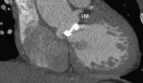
ECG-gated or -triggered CT angiograms were selected from our PACS archive based on the presence of prosthetic heart valves. The best systolic and diastolic axial reconstructions were selected for coronary assessment. Each present coronary segment was scored for the presence of valve-related artefacts prohibiting coronary artery assessment. Scoring was performed in consensus by two observers.
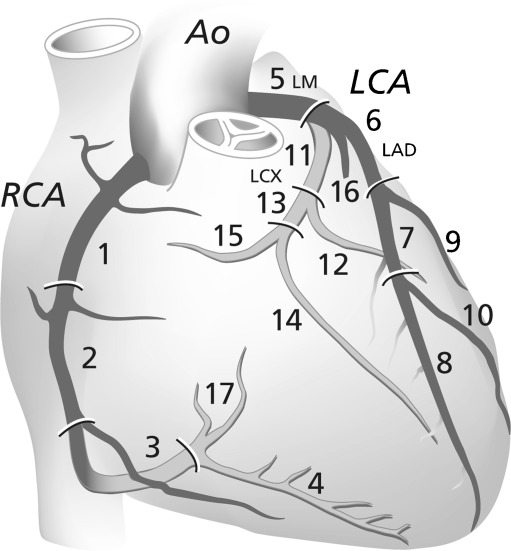
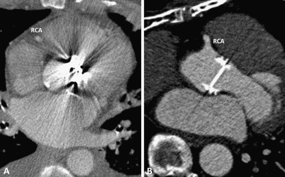
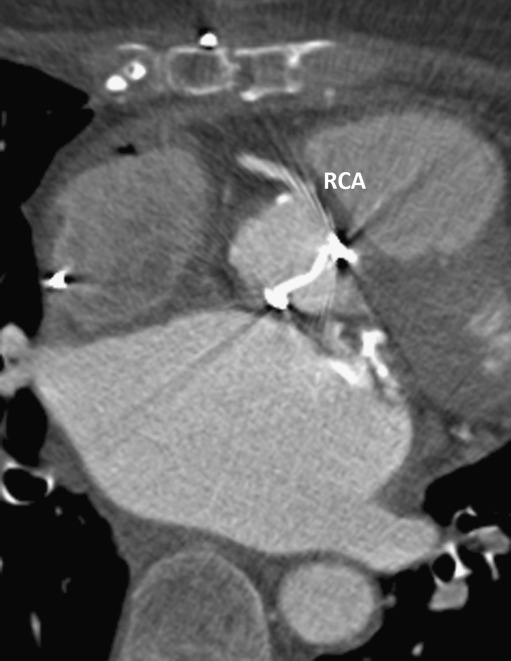
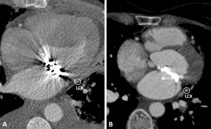
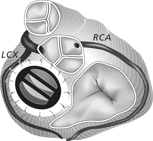
Eighty-two CT angiograms were performed on a 64-slice (n = 27) or 256-slice (n = 55) multidetector CT. Eighty-nine valves and five annuloplasty rings were present. Forty-three out of 1160 (3.7%) present coronary artery segments were non-diagnostic due to valve artefacts (14/82 patients). Valve artefacts were located in right coronary artery (15/43; 35%), left anterior descending artery (2/43; 5%), circumflex artery (14/43; 32%) and marginal obtuse (12/43; 28%) segments. All cobalt-chrome containing valves caused artefacts prohibiting coronary assessment. Biological and titanium-containing valves did not cause artefacts except for three specific valve types.
Most commonly implanted prosthetic heart valves do not hamper coronary assessment on multidetector CT. Cobalt-chrome containing prosthetic heart valves preclude complete coronary artery assessment because of severe valve artefacts.
• Most commonly implanted prosthetic heart valves do not hamper coronary artery assessment • Prosthetic heart valve composition determines the occurrence of prosthetic heart valve-related artefacts • Björk-Shiley and Sorin tilting disc valves preclude diagnostic coronary artery segment assessment.
人工心脏瓣膜患者可能需要评估冠状动脉疾病。我们评估了多层 CT 是否会因瓣膜伪影而妨碍冠状动脉评估。
根据是否存在人工心脏瓣膜,从我们的 PACS 档案中选择 ECG 门控或触发 CT 血管造影。选择最佳的收缩期和舒张期轴向重建进行冠状动脉评估。对每个存在的冠状动脉节段进行评分,以确定是否存在妨碍冠状动脉评估的瓣膜相关伪影。评分由两名观察者进行共识评估。
在 64 层(n=27)或 256 层(n=55)多层 CT 上进行了 82 次 CT 血管造影。存在 89 个瓣膜和 5 个瓣环成形环。由于瓣膜伪影,1160 个存在的冠状动脉节段中有 43 个(14/82 例患者)无法诊断。瓣膜伪影位于右冠状动脉(15/43;35%)、左前降支(2/43;5%)、回旋支(14/43;32%)和边缘钝缘支(12/43;28%)。所有含钴铬的瓣膜都会引起妨碍冠状动脉评估的伪影。生物和钛合金瓣膜不会引起伪影,除了三种特定的瓣膜类型。
大多数植入的人工心脏瓣膜不会在多层 CT 上妨碍冠状动脉评估。含钴铬的人工心脏瓣膜会因严重的瓣膜伪影而排除完整的冠状动脉评估。
大多数植入的人工心脏瓣膜不会妨碍冠状动脉评估。
人工心脏瓣膜的组成决定了人工心脏瓣膜相关伪影的发生。
Björk-Shiley 和 Sorin 倾斜碟阀排除了诊断性冠状动脉节段评估。