Department of Microbiology and Molecular Medicine, University Hospital and Medical School of Geneva, Geneva, Switzerland.
PLoS Pathog. 2011 Dec;7(12):e1002456. doi: 10.1371/journal.ppat.1002456. Epub 2011 Dec 15.
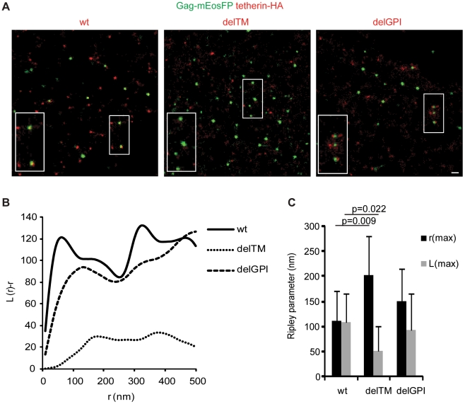
Virus assembly and interaction with host-cell proteins occur at length scales below the diffraction limit of visible light. Novel super-resolution microscopy techniques achieve nanometer resolution of fluorescently labeled molecules. The cellular restriction factor tetherin (also known as CD317, BST-2 or HM1.24) inhibits the release of human immunodeficiency virus 1 (HIV-1) through direct incorporation into viral membranes and is counteracted by the HIV-1 protein Vpu. For super-resolution analysis of HIV-1 and tetherin interactions, we established fluorescence labeling of HIV-1 proteins and tetherin that preserved HIV-1 particle formation and Vpu-dependent restriction, respectively. Multicolor super-resolution microscopy revealed important structural features of individual HIV-1 virions, virus assembly sites and their interaction with tetherin at the plasma membrane. Tetherin localization to micro-domains was dependent on both tetherin membrane anchors. Tetherin clusters containing on average 4 to 7 tetherin dimers were visualized at HIV-1 assembly sites. Combined biochemical and super-resolution analysis revealed that extended tetherin dimers incorporate both N-termini into assembling virus particles and restrict HIV-1 release. Neither tetherin domains nor HIV-1 assembly sites showed enrichment of the raft marker GM1. Together, our super-resolution microscopy analysis of HIV-1 interactions with tetherin provides new insights into the mechanism of tetherin-mediated HIV-1 restriction and paves the way for future studies of virus-host interactions.
病毒组装和与宿主细胞蛋白的相互作用发生在可见光衍射极限以下的长度尺度上。新型超分辨率显微镜技术实现了荧光标记分子的纳米级分辨率。细胞限制因子 tetherin(也称为 CD317、BST-2 或 HM1.24)通过直接掺入病毒膜来抑制人类免疫缺陷病毒 1(HIV-1)的释放,并且 HIV-1 蛋白 Vpu 会拮抗 tetherin。为了对 HIV-1 和 tetherin 相互作用进行超分辨率分析,我们建立了 HIV-1 蛋白和 tetherin 的荧光标记,分别保留了 HIV-1 颗粒形成和 Vpu 依赖性限制。多色超分辨率显微镜揭示了单个 HIV-1 病毒粒子、病毒组装位点及其与质膜上 tetherin 的相互作用的重要结构特征。tetherin 的定位依赖于质膜锚定的 tetherin 膜锚定。在 HIV-1 组装位点处可视化到含有平均 4 到 7 个 tetherin 二聚体的 tetherin 簇。结合生化和超分辨率分析表明,扩展的 tetherin 二聚体将两个 N 末端都纳入组装的病毒颗粒中,并限制 HIV-1 的释放。tetherin 结构域和 HIV-1 组装位点均未显示筏标记 GM1 的富集。总之,我们对 HIV-1 与 tetherin 相互作用的超分辨率显微镜分析提供了对 tetherin 介导的 HIV-1 限制机制的新见解,并为未来研究病毒-宿主相互作用铺平了道路。