Section of Cell Biology II, Netherlands Cancer Institute, Amsterdam, The Netherlands.
PLoS Pathog. 2011 Dec;7(12):e1002449. doi: 10.1371/journal.ppat.1002449. Epub 2011 Dec 22.
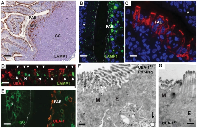
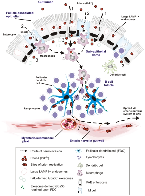
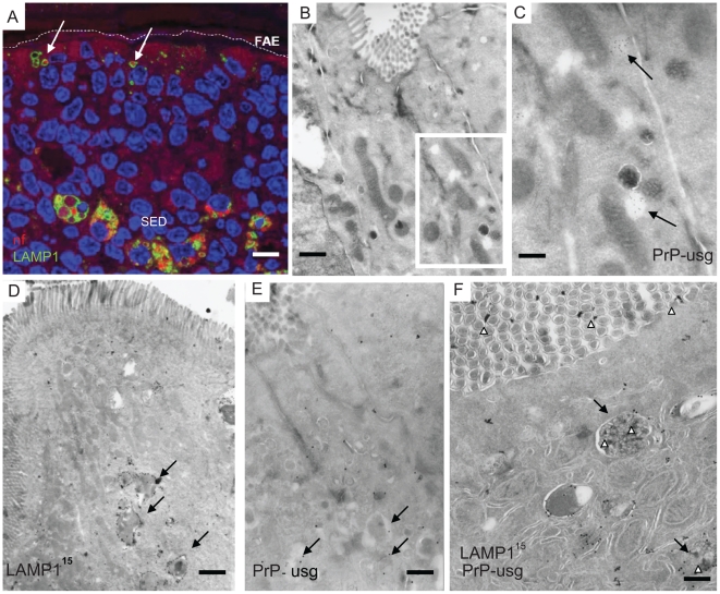
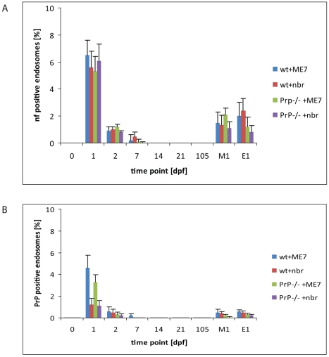
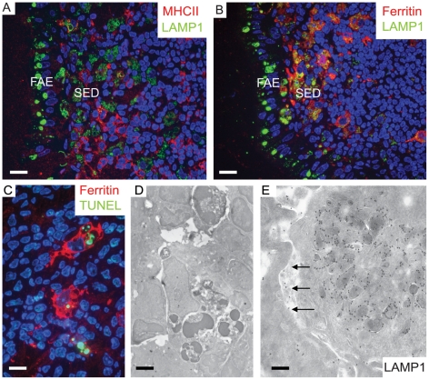
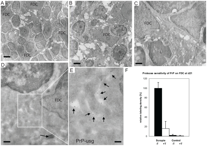
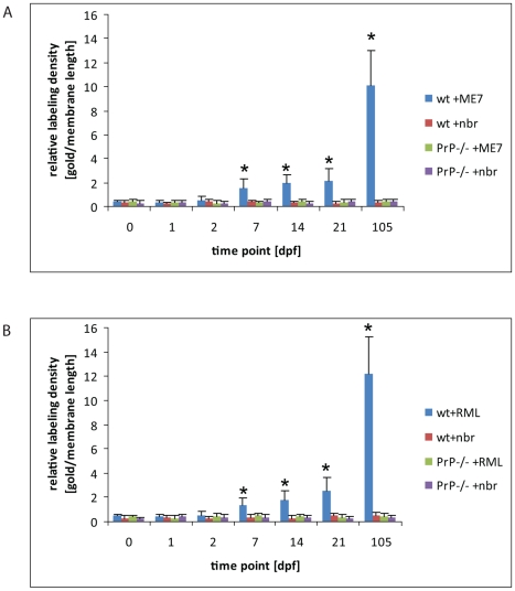
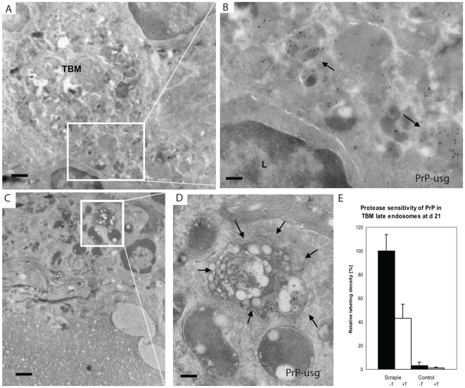
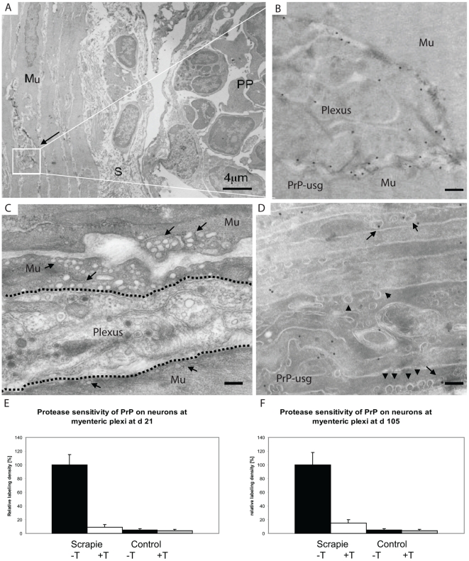
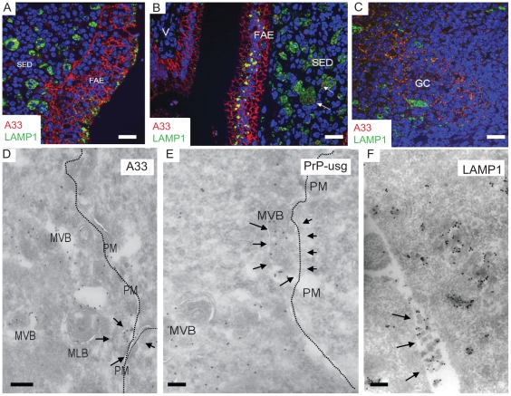
After oral exposure, prions are thought to enter Peyer's patches via M cells and accumulate first upon follicular dendritic cells (FDCs) before spreading to the nervous system. How prions are actually initially acquired from the gut lumen is not known. Using high-resolution immunofluorescence and cryo-immunogold electron microscopy, we report the trafficking of the prion protein (PrP) toward Peyer's patches of wild-type and PrP-deficient mice. PrP was transiently detectable at 1 day post feeding (dpf) within large multivesicular LAMP1-positive endosomes of enterocytes in the follicle-associated epithelium (FAE) and at much lower levels within M cells. Subsequently, PrP was detected on vesicles in the late endosomal compartments of macrophages in the subepithelial dome. At 7-21 dpf, increased PrP labelling was observed on the plasma membranes of FDCs in germinal centres of Peyer's patches from wild-type mice only, identifying FDCs as the first sites of PrP conversion and replication. Detection of PrP on extracellular vesicles displaying FAE enterocyte-derived A33 protein implied transport towards FDCs in association with FAE-derived vesicles. By 21 dpf, PrP was observed on the plasma membranes of neurons within neighbouring myenteric plexi. Together, these data identify a novel potential M cell-independent mechanism for prion transport, mediated by FAE enterocytes, which acts to initiate conversion and replication upon FDCs and subsequent infection of enteric nerves.
经口暴露后,朊病毒被认为通过 M 细胞进入派尔集合淋巴结,并首先在滤泡树突状细胞(FDC)上积累,然后传播到神经系统。目前尚不清楚朊病毒实际上是如何从肠腔中最初获得的。我们使用高分辨率免疫荧光和冷冻免疫金电子显微镜技术,报告了野生型和 PrP 缺陷型小鼠派尔集合淋巴结中朊病毒蛋白(PrP)的转运。在喂食后 1 天(dpf),PrP 可在滤泡相关上皮(FAE)中的肠细胞大的多泡 LAMP1 阳性内体中短暂检测到,在 M 细胞中检测到的水平要低得多。随后,在黏膜下层穹窿中的巨噬细胞的晚期内体隔室中的小泡中检测到 PrP。在 7-21 dpf 时,仅在野生型小鼠的派尔集合淋巴结生发中心的 FDC 上观察到 PrP 标记增加,这表明 FDC 是 PrP 转化和复制的第一个部位。在显示 FAE 肠细胞衍生的 A33 蛋白的细胞外小泡上检测到 PrP,这表明它与 FAE 衍生的小泡一起向 FDC 运输。在 21 dpf 时,观察到邻近肌间神经丛中的神经元的质膜上存在 PrP。总之,这些数据确定了一种新型潜在的 M 细胞非依赖性朊病毒转运机制,由 FAE 肠细胞介导,该机制在 FDC 上发挥作用,从而在 FDC 上引发转化和复制,随后感染肠神经。