• 文献检索
  • 文档翻译
  • 深度研究
  • 学术资讯
  • Suppr Zotero 插件Zotero 插件
  • 邀请有礼
  • 套餐&价格
  • 历史记录
应用&插件
Suppr Zotero 插件Zotero 插件浏览器插件Mac 客户端Windows 客户端微信小程序
定价
高级版会员购买积分包购买API积分包
服务
文献检索文档翻译深度研究API 文档MCP 服务
关于我们
关于 Suppr公司介绍联系我们用户协议隐私条款
关注我们

Suppr 超能文献

核心技术专利:CN118964589B侵权必究
粤ICP备2023148730 号-1Suppr @ 2026

文献检索

告别复杂PubMed语法,用中文像聊天一样搜索,搜遍4000万医学文献。AI智能推荐,让科研检索更轻松。

立即免费搜索

文件翻译

保留排版,准确专业,支持PDF/Word/PPT等文件格式,支持 12+语言互译。

免费翻译文档

深度研究

AI帮你快速写综述,25分钟生成高质量综述,智能提取关键信息,辅助科研写作。

立即免费体验

M 细胞耗竭阻断经口朊病毒病发病机制。

M cell-depletion blocks oral prion disease pathogenesis.

机构信息

The Roslin Institute and Royal (Dick) School of Veterinary Sciences, University of Edinburgh, Edinburgh, UK.

出版信息

Mucosal Immunol. 2012 Mar;5(2):216-25. doi: 10.1038/mi.2011.68. Epub 2012 Feb 1.

DOI:10.1038/mi.2011.68
PMID:22294048
原文链接:https://pmc.ncbi.nlm.nih.gov/articles/PMC3282432/
Abstract

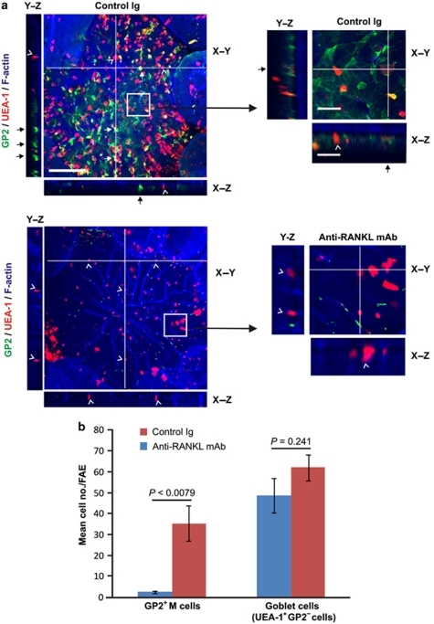
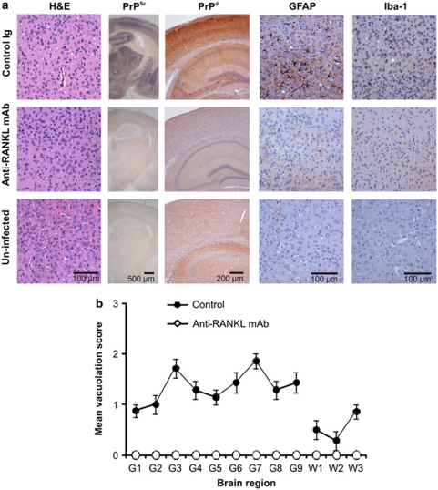
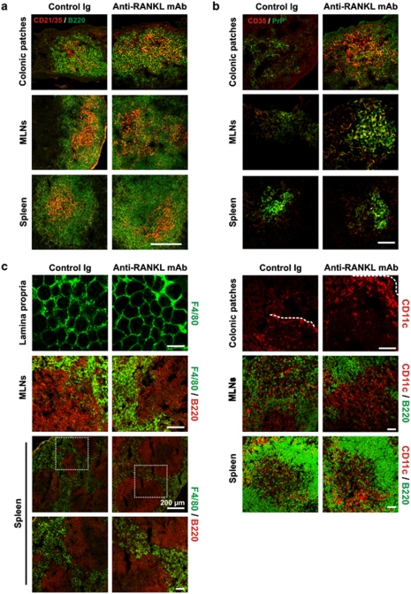
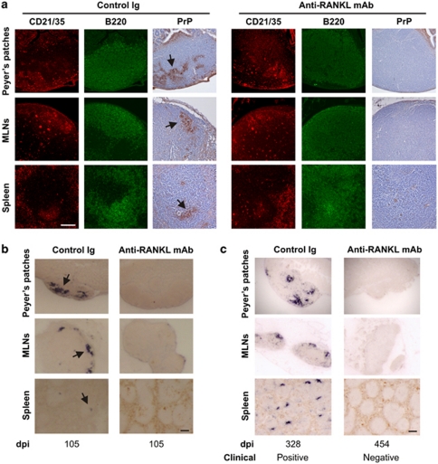
Many prion diseases are orally acquired. Our data show that after oral exposure, early prion replication upon follicular dendritic cells (FDC) in Peyer's patches is obligatory for the efficient spread of disease to the brain (termed neuroinvasion). For prions to replicate on FDC within Peyer's patches after ingestion of a contaminated meal, they must first cross the gut epithelium. However, the mechanism through which prions are conveyed into Peyer's patches is uncertain. Within the follicle-associated epithelium overlying Peyer's patches are microfold cells (M cells), unique epithelial cells specialized for the transcytosis of particles. We show that following M cell-depletion, early prion accumulation upon FDC in Peyer's patches is blocked. Furthermore, in the absence of M cells at the time of oral exposure, neuroinvasion and disease development are likewise blocked. These data suggest M cells are important sites of prion uptake from the gut lumen into Peyer's patches.

摘要

许多朊病毒疾病是经口获得的。我们的数据表明,在口服暴露后,滤泡树突状细胞(FDC)在派尔集合淋巴结中的早期朊病毒复制对于疾病向大脑的有效传播(称为神经入侵)是必需的。为了使摄入污染食物后的 FDC 在派尔集合淋巴结中复制朊病毒,它们必须首先穿过肠道上皮。然而,朊病毒被递送到派尔集合淋巴结的机制尚不确定。在派尔集合淋巴结上的滤泡相关上皮内是微褶皱细胞(M 细胞),是专门用于颗粒胞吞作用的独特上皮细胞。我们发现,在 M 细胞耗竭后,派尔集合淋巴结中 FDC 上的早期朊病毒积累被阻断。此外,在口服暴露时不存在 M 细胞的情况下,神经入侵和疾病发展也同样被阻断。这些数据表明 M 细胞是从肠道腔摄取朊病毒进入派尔集合淋巴结的重要部位。

https://cdn.ncbi.nlm.nih.gov/pmc/blobs/90b7/3282432/66a01bb78b06/mi201168f4.jpg
https://cdn.ncbi.nlm.nih.gov/pmc/blobs/90b7/3282432/2e63fa31daf2/mi201168f1.jpg
https://cdn.ncbi.nlm.nih.gov/pmc/blobs/90b7/3282432/3604b0c6d273/mi201168f2.jpg
https://cdn.ncbi.nlm.nih.gov/pmc/blobs/90b7/3282432/36d63cc4c42c/mi201168f3.jpg
https://cdn.ncbi.nlm.nih.gov/pmc/blobs/90b7/3282432/66a01bb78b06/mi201168f4.jpg

相似文献

1
M cell-depletion blocks oral prion disease pathogenesis.M 细胞耗竭阻断经口朊病毒病发病机制。
Mucosal Immunol. 2012 Mar;5(2):216-25. doi: 10.1038/mi.2011.68. Epub 2012 Feb 1.
2
Oral Prion Neuroinvasion Occurs Independently of PrP Expression in the Gut Epithelium.口腔朊病毒神经入侵独立于肠道上皮细胞中的 PrP 表达。
J Virol. 2018 Sep 12;92(19). doi: 10.1128/JVI.01010-18. Print 2018 Oct 1.
3
Oral Prion Disease Pathogenesis Is Impeded in the Specific Absence of CXCR5-Expressing Dendritic Cells.在特异性缺失表达CXCR5的树突状细胞的情况下,口腔朊病毒病的发病机制受到阻碍。
J Virol. 2017 Apr 28;91(10). doi: 10.1128/JVI.00124-17. Print 2017 May 15.
4
Prion uptake in the gut: identification of the first uptake and replication sites.朊病毒在肠道中的摄取:首次摄取和复制部位的鉴定。
PLoS Pathog. 2011 Dec;7(12):e1002449. doi: 10.1371/journal.ppat.1002449. Epub 2011 Dec 22.
5
Increased Abundance of M Cells in the Gut Epithelium Dramatically Enhances Oral Prion Disease Susceptibility.肠道上皮中M细胞数量增加显著增强口腔朊病毒病易感性。
PLoS Pathog. 2016 Dec 14;12(12):e1006075. doi: 10.1371/journal.ppat.1006075. eCollection 2016 Dec.
6
Orally administered prion protein is incorporated by m cells and spreads into lymphoid tissues with macrophages in prion protein knockout mice.经口给予的朊病毒蛋白被 M 细胞摄取,并在朊病毒蛋白敲除小鼠中与巨噬细胞一起扩散到淋巴组织中。
Am J Pathol. 2011 Sep;179(3):1301-9. doi: 10.1016/j.ajpath.2011.05.058. Epub 2011 Jul 18.
7
Neuroimmune connections in jejunal and ileal Peyer's patches at various bovine ages: potential sites for prion neuroinvasion.不同牛龄空肠和回肠派尔集合淋巴结中的神经免疫连接:朊病毒神经侵袭的潜在部位
Cell Tissue Res. 2007 Jul;329(1):35-44. doi: 10.1007/s00441-007-0396-4. Epub 2007 Apr 4.
8
Role of the GALT in scrapie agent neuroinvasion from the intestine.肠道相关淋巴组织(GALT)在瘙痒病病原体从肠道向神经侵袭过程中的作用。
J Immunol. 2007 Mar 15;178(6):3757-66. doi: 10.4049/jimmunol.178.6.3757.
9
The Gut-Associated Lymphoid Tissues in the Small Intestine, Not the Large Intestine, Play a Major Role in Oral Prion Disease Pathogenesis.小肠而非大肠中的肠道相关淋巴组织在经口感染朊病毒疾病发病机制中起主要作用。
J Virol. 2015 Sep;89(18):9532-47. doi: 10.1128/JVI.01544-15. Epub 2015 Jul 8.
10
c-Rel is dispensable for the differentiation and functional maturation of M cells in the follicle-associated epithelium.c-Rel对于滤泡相关上皮中M细胞的分化和功能成熟并非必需。
Immunobiology. 2017 Feb;222(2):316-326. doi: 10.1016/j.imbio.2016.09.008. Epub 2016 Sep 18.

引用本文的文献

1
Mediators linking gut microbiota and sporadic Creutzfeldt-Jakob disease: a Mendelian randomization study.连接肠道微生物群与散发性克雅氏病的介质:一项孟德尔随机化研究
AMB Express. 2025 Aug 18;15(1):120. doi: 10.1186/s13568-025-01932-3.
2
Acute LPS exposure enhances susceptibility to peripheral prion infection.急性接触脂多糖会增加对外周朊病毒感染的易感性。
Sci Rep. 2025 Mar 21;15(1):9754. doi: 10.1038/s41598-025-94003-3.
3
Food antigens suppress small intestinal tumorigenesis.食物抗原抑制小肠肿瘤发生。

本文引用的文献

1
Follicular dendritic cell-specific prion protein (PrP) expression alone is sufficient to sustain prion infection in the spleen.滤泡树突状细胞特异性朊病毒蛋白 (PrP) 的单独表达足以维持脾脏中的朊病毒感染。
PLoS Pathog. 2011 Dec;7(12):e1002402. doi: 10.1371/journal.ppat.1002402. Epub 2011 Dec 1.
2
Convergent and divergent development among M cell lineages in mouse mucosal epithelium.M 细胞谱系在小鼠黏膜上皮中的趋同和分歧发育。
J Immunol. 2011 Nov 15;187(10):5277-85. doi: 10.4049/jimmunol.1102077. Epub 2011 Oct 7.
3
Distinct developmental requirements for isolated lymphoid follicle formation in the small and large intestine: RANKL is essential only in the small intestine.
Front Immunol. 2024 Sep 18;15:1373766. doi: 10.3389/fimmu.2024.1373766. eCollection 2024.
4
Perspectives on CRISPR Genome Editing to Prevent Prion Diseases in High-Risk Individuals.关于CRISPR基因编辑预防高危个体朊病毒疾病的观点。
Biomedicines. 2024 Aug 1;12(8):1725. doi: 10.3390/biomedicines12081725.
5
Differential role of M cells in enteroid infection by subsp. and serovar Typhimurium.肠类器官感染 亚血清型 和 鼠伤寒血清型中 M 细胞的差异作用。
Front Cell Infect Microbiol. 2024 Jul 8;14:1416537. doi: 10.3389/fcimb.2024.1416537. eCollection 2024.
6
Deciphering the M-cell niche: insights from mouse models on how microfold cells "know" where they are needed.解析 M 细胞生态位:从小鼠模型中了解微皱褶细胞如何“知道”它们需要出现在哪里。
Front Immunol. 2024 May 28;15:1400739. doi: 10.3389/fimmu.2024.1400739. eCollection 2024.
7
Dysbiosis of the gut microbiota and its effect on α-synuclein and prion protein misfolding: consequences for neurodegeneration.肠道微生物群失调及其对α-突触核蛋白和朊病毒蛋白错误折叠的影响:对神经退行性变的影响。
Front Cell Infect Microbiol. 2024 Feb 16;14:1348279. doi: 10.3389/fcimb.2024.1348279. eCollection 2024.
8
Strain-Specific Targeting and Destruction of Cells by Prions.朊病毒对特定毒株细胞的靶向作用与破坏
Biology (Basel). 2024 Jan 20;13(1):57. doi: 10.3390/biology13010057.
9
Inhibition of microfold cells ameliorates early pathological phenotypes by modulating microglial functions in Alzheimer's disease mouse model.抑制微小褶皱细胞通过调节阿尔茨海默病小鼠模型中的小胶质细胞功能改善早期病理表型。
J Neuroinflammation. 2023 Nov 27;20(1):282. doi: 10.1186/s12974-023-02966-9.
10
Creutzfeldt-Jakob Disease: Alterations of Gut Microbiota.克雅氏病:肠道微生物群的改变
Front Neurol. 2022 Apr 15;13:832599. doi: 10.3389/fneur.2022.832599. eCollection 2022.
在小肠和大肠中形成孤立淋巴滤泡的不同发育要求:RANKL 在小肠中是必需的。
Am J Pathol. 2011 Oct;179(4):1861-71. doi: 10.1016/j.ajpath.2011.06.004. Epub 2011 Aug 18.
4
Orally administered prion protein is incorporated by m cells and spreads into lymphoid tissues with macrophages in prion protein knockout mice.经口给予的朊病毒蛋白被 M 细胞摄取,并在朊病毒蛋白敲除小鼠中与巨噬细胞一起扩散到淋巴组织中。
Am J Pathol. 2011 Sep;179(3):1301-9. doi: 10.1016/j.ajpath.2011.05.058. Epub 2011 Jul 18.
5
Expression of mesenchyme-specific gene signatures by follicular dendritic cells: insights from the meta-analysis of microarray data from multiple mouse cell populations.滤泡树突状细胞表达间充质特异性基因特征:来自多个小鼠细胞群体的基因芯片数据分析的荟萃分析。
Immunology. 2011 Aug;133(4):482-98. doi: 10.1111/j.1365-2567.2011.03461.x. Epub 2011 Jun 3.
6
CCR6hiCD11c(int) B cells promote M-cell differentiation in Peyer's patch.CCR6hiCD11c(int) B 细胞促进派氏集合淋巴结中 M 细胞的分化。
Int Immunol. 2011 Apr;23(4):261-9. doi: 10.1093/intimm/dxq478. Epub 2011 Mar 21.
7
Murine cecal patch M cells transport infectious prions in vivo.鼠盲肠斑片 M 细胞在体内转运感染性朊病毒。
J Infect Dis. 2010 Dec 15;202(12):1916-9. doi: 10.1086/657415. Epub 2010 Nov 4.
8
Meta-analysis of lineage-specific gene expression signatures in mouse leukocyte populations.小鼠白细胞群体谱系特异性基因表达特征的荟萃分析。
Immunobiology. 2010 Sep-Oct;215(9-10):724-36. doi: 10.1016/j.imbio.2010.05.012. Epub 2010 Jun 4.
9
M-Sec promotes membrane nanotube formation by interacting with Ral and the exocyst complex.M-Sec通过与Ral和外排体复合物相互作用来促进膜纳米管的形成。
Nat Cell Biol. 2009 Dec;11(12):1427-32. doi: 10.1038/ncb1990. Epub 2009 Nov 22.
10
Uptake through glycoprotein 2 of FimH(+) bacteria by M cells initiates mucosal immune response.FimH(+)细菌通过糖蛋白 2 被 M 细胞摄取,从而引发黏膜免疫应答。
Nature. 2009 Nov 12;462(7270):226-30. doi: 10.1038/nature08529.