• 文献检索
  • 文档翻译
  • 深度研究
  • 学术资讯
  • Suppr Zotero 插件Zotero 插件
  • 邀请有礼
  • 套餐&价格
  • 历史记录
应用&插件
Suppr Zotero 插件Zotero 插件浏览器插件Mac 客户端Windows 客户端微信小程序
定价
高级版会员购买积分包购买API积分包
服务
文献检索文档翻译深度研究API 文档MCP 服务
关于我们
关于 Suppr公司介绍联系我们用户协议隐私条款
关注我们

Suppr 超能文献

核心技术专利:CN118964589B侵权必究
粤ICP备2023148730 号-1Suppr @ 2026

文献检索

告别复杂PubMed语法,用中文像聊天一样搜索,搜遍4000万医学文献。AI智能推荐,让科研检索更轻松。

立即免费搜索

文件翻译

保留排版,准确专业,支持PDF/Word/PPT等文件格式,支持 12+语言互译。

免费翻译文档

深度研究

AI帮你快速写综述,25分钟生成高质量综述,智能提取关键信息,辅助科研写作。

立即免费体验

C 臂透视模式下吸收剂量的比较研究。

A Study to Compare the Radiation Absorbed Dose of the C-arm Fluoroscopic Modes.

机构信息

Department of Anesthesiology and Pain Medicine, Konkuk University Medical Center, Seoul, Korea.

出版信息

Korean J Pain. 2011 Dec;24(4):199-204. doi: 10.3344/kjp.2011.24.4.199. Epub 2011 Nov 30.

DOI:10.3344/kjp.2011.24.4.199
PMID:22220241
原文链接:https://pmc.ncbi.nlm.nih.gov/articles/PMC3248583/
Abstract

BACKGROUND

Although many clinicians know about the reducing effects of the pulsed and low-dose modes for fluoroscopic radiation when performing interventional procedures, few studies have quantified the reduction of radiation-absorbed doses (RADs). The aim of this study is to compare how much the RADs from a fluoroscopy are reduced according to the C-arm fluoroscopic modes used.

METHODS

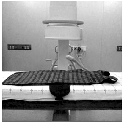
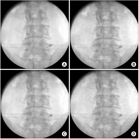
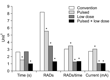
We measured the RADs in the C-arm fluoroscopic modes including 'conventional mode', 'pulsed mode', 'low-dose mode', and 'pulsed + low-dose mode'. Clinical imaging conditions were simulated using a lead apron instead of a patient. According to each mode, one experimenter radiographed the lead apron, which was on the table, consecutively 5 times on the AP views. We regarded this as one set and a total of 10 sets were done according to each mode. Cumulative exposure time, RADs, peak X-ray energy, and current, which were viewed on the monitor, were recorded.

RESULTS

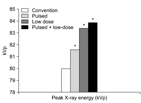
Pulsed, low-dose, and pulsed + low-dose modes showed significantly decreased RADs by 32%, 57%, and 83% compared to the conventional mode. The mean cumulative exposure time was significantly lower in the pulsed and pulsed + low-dose modes than in the conventional mode. All modes had pretty much the same peak X-ray energy. The mean current was significantly lower in the low-dose and pulsed + low-dose modes than in the conventional mode.

CONCLUSIONS

The use of the pulsed and low-dose modes together significantly reduced the RADs compared to the conventional mode. Therefore, the proper use of the fluoroscopy and its C-arm modes will reduce the radiation exposure of patients and clinicians.

摘要

背景

虽然许多临床医生在进行介入性手术时都知道脉冲和低剂量模式可降低透视辐射的放射剂量,但很少有研究量化放射吸收剂量(RAD)的减少。本研究旨在比较使用不同 C 臂透视模式时,透视的 RAD 减少了多少。

方法

我们测量了 C 臂透视模式下的 RAD,包括“常规模式”、“脉冲模式”、“低剂量模式”和“脉冲+低剂量模式”。使用铅围裙代替患者模拟临床成像条件。根据每种模式,一名实验人员连续五次对放在桌上的铅围裙进行 AP 视图拍摄。我们将这视为一组,每种模式总共进行 10 组。记录在监视器上查看的累积曝光时间、RAD、峰值 X 射线能量和电流。

结果

与常规模式相比,脉冲、低剂量和脉冲+低剂量模式的 RAD 分别显著降低了 32%、57%和 83%。与常规模式相比,脉冲和脉冲+低剂量模式的平均累积曝光时间明显更低。所有模式的峰值 X 射线能量几乎相同。低剂量和脉冲+低剂量模式的平均电流明显低于常规模式。

结论

与常规模式相比,同时使用脉冲和低剂量模式可显著降低 RAD。因此,正确使用透视及其 C 臂模式将降低患者和临床医生的辐射暴露。

https://cdn.ncbi.nlm.nih.gov/pmc/blobs/8339/3248583/9dd26228c4fb/kjpain-24-199-g004.jpg
https://cdn.ncbi.nlm.nih.gov/pmc/blobs/8339/3248583/93ff0fc580bc/kjpain-24-199-g001.jpg
https://cdn.ncbi.nlm.nih.gov/pmc/blobs/8339/3248583/27426346aaf8/kjpain-24-199-g002.jpg
https://cdn.ncbi.nlm.nih.gov/pmc/blobs/8339/3248583/6dd98f74156e/kjpain-24-199-g003.jpg
https://cdn.ncbi.nlm.nih.gov/pmc/blobs/8339/3248583/9dd26228c4fb/kjpain-24-199-g004.jpg

相似文献

1
A Study to Compare the Radiation Absorbed Dose of the C-arm Fluoroscopic Modes.C 臂透视模式下吸收剂量的比较研究。
Korean J Pain. 2011 Dec;24(4):199-204. doi: 10.3344/kjp.2011.24.4.199. Epub 2011 Nov 30.
2
Reduction of radiation exposure while maintaining high-quality fluoroscopic images during interventional cardiology using novel x-ray tube technology with extra beam filtering.在介入心脏病学中,使用具有额外光束过滤功能的新型X射线管技术,在保持高质量荧光透视图像的同时减少辐射暴露。
Circulation. 1994 Jun;89(6):2710-4. doi: 10.1161/01.cir.89.6.2710.
3
Reduction in average fluoroscopic exposure times for interventional spinal procedures through the use of pulsed and low-dose image settings.通过使用脉冲和低剂量图像设置,降低介入性脊柱手术的平均透视曝光时间。
Am J Phys Med Rehabil. 2011 Nov;90(11):908-12. doi: 10.1097/PHM.0b013e318228c9dd.
4
[Reduction of radiation exposure by the use of fluoroscopic guidance in transpedicular instrumentation].[在经椎弓根器械植入术中使用透视引导减少辐射暴露]
Acta Chir Orthop Traumatol Cech. 2011;78(5):447-50.
5
[Initial experiences with pulsed fluoroscopy on a multifunctional fluoroscopic unit].[多功能荧光透视设备上脉冲透视的初步经验]
Rofo. 1996 Nov;165(5):475-9. doi: 10.1055/s-2007-1015793.
6
Radiation exposure during continuous and pulsed fluoroscopy.连续透视和脉冲透视中的辐射暴露。
J Endourol. 2013 Mar;27(3):384-8. doi: 10.1089/end.2012.0213. Epub 2012 Nov 28.
7
Reducing Radiation Exposure in Lumbar Transforaminal Epidural Steroid Injections with Pulsed Fluoroscopy: A Randomized, Double-blind, Controlled Clinical Trial.应用脉冲透视降低腰椎经椎间孔硬膜外类固醇注射的辐射暴露:一项随机、双盲、对照临床试验。
Pain Physician. 2018 Jan;21(1):53-60.
8
Patient skin dose in cardiac interventional procedures: conventional fluoroscopy versus pulsed fluoroscopy.心脏介入手术中的患者皮肤剂量:传统透视与脉冲透视对比
Catheter Cardiovasc Interv. 2007 Jan;69(1):115-21; discussion 122. doi: 10.1002/ccd.20961.
9
Reduction of radiation dose in pediatric patients using pulsed fluoroscopy.使用脉冲透视法降低儿科患者的辐射剂量。
AJR Am J Roentgenol. 1996 Nov;167(5):1247-53. doi: 10.2214/ajr.167.5.8911190.
10
Radiation Exposure to the Surgeon and Patient During a Fluoroscopic Procedure: How High Is the Exposure Dose? A Cadaveric Study.透视检查过程中外科医生和患者所受的辐射暴露:暴露剂量有多高?一项尸体研究。
Spine (Phila Pa 1976). 2016 Aug 1;41(15):1254-1260. doi: 10.1097/BRS.0000000000001542.

引用本文的文献

1
Intraoperative radiation exposure in a level 1 trauma centre orthopedic operating room.一级创伤中心骨科手术室的术中辐射暴露
Can J Surg. 2025 Jun 10;68(3):E235-E241. doi: 10.1503/cjs.003824. Print 2025 May-Jun.
2
Optimizing DSA parameters for enhanced radiation safety in interventional surgery.优化数字减影血管造影(DSA)参数以提高介入手术中的辐射安全性。
Front Public Health. 2025 Feb 21;13:1531240. doi: 10.3389/fpubh.2025.1531240. eCollection 2025.
3
Radiation safety for pain physicians: principles and recommendations.疼痛科医生的辐射安全:原则与建议

本文引用的文献

1
Reduction in average fluoroscopic exposure times for interventional spinal procedures through the use of pulsed and low-dose image settings.通过使用脉冲和低剂量图像设置,降低介入性脊柱手术的平均透视曝光时间。
Am J Phys Med Rehabil. 2011 Nov;90(11):908-12. doi: 10.1097/PHM.0b013e318228c9dd.
2
Radiation exposure to physicians during interventional pain procedures.介入性疼痛治疗程序中医生的辐射暴露。
Korean J Pain. 2010 Mar;23(1):24-7. doi: 10.3344/kjp.2010.23.1.24. Epub 2010 Mar 10.
3
Occupational radiation protection in interventional radiology: a joint guideline of the Cardiovascular and Interventional Radiology Society of Europe and the Society of Interventional Radiology.
Korean J Pain. 2022 Apr 1;35(2):129-139. doi: 10.3344/kjp.2022.35.2.129.
4
[Intraoperative imaging of children and adolescents, for selected fractures and in follow-up after conservative and operative treatment : Part 2 of the results of a nationwide online survey of the Pediatric Traumatology Section of the German Trauma Society].[儿童和青少年术中成像,用于特定骨折以及保守和手术治疗后的随访:德国创伤学会儿科创伤学分会全国在线调查结果的第2部分]
Unfallchirurgie (Heidelb). 2023 Jan;126(1):42-54. doi: 10.1007/s00113-021-01114-3. Epub 2021 Dec 16.
5
Interventional Radiology Suite: A Primer for Trainees.介入放射学套件:实习生入门指南。
J Clin Med. 2019 Aug 30;8(9):1347. doi: 10.3390/jcm8091347.
6
Radiation dose to the operator during fluoroscopically guided spine procedures.透视引导下脊柱手术期间术者所接受的辐射剂量。
Neuroradiology. 2017 Sep;59(9):885-892. doi: 10.1007/s00234-017-1868-1. Epub 2017 Jul 18.
7
The radiation safety education and the pain physicians' efforts to reduce radiation exposure.辐射安全教育以及疼痛科医生为减少辐射暴露所做的努力。
Korean J Pain. 2017 Apr;30(2):104-115. doi: 10.3344/kjp.2017.30.2.104. Epub 2017 Mar 31.
8
Use of C-Arm to Assess Reduction of Zygomatic Complex Fractures: A Comparative Study.使用C型臂评估颧骨复合体骨折的复位情况:一项对比研究。
Craniomaxillofac Trauma Reconstr. 2017 Mar;10(1):35-43. doi: 10.1055/s-0036-1592085. Epub 2016 Nov 7.
9
The Radiation Exposure of Radiographer Related to the Location in C-arm Fluoroscopy-guided Pain Interventions.C 臂透视引导下疼痛介入放射技师的辐射暴露与位置的关系。
Korean J Pain. 2014 Apr;27(2):162-7. doi: 10.3344/kjp.2014.27.2.162. Epub 2014 Mar 28.
10
How Effective Are Radiation Reducing Gloves in C-arm Fluoroscopy-guided Pain Interventions?辐射降低手套在 C 臂透视引导下疼痛介入中的效果如何?
Korean J Pain. 2014 Apr;27(2):145-51. doi: 10.3344/kjp.2014.27.2.145. Epub 2014 Mar 28.
介入放射学中的职业辐射防护:欧洲心血管和介入放射学会与介入放射学会联合指南
Cardiovasc Intervent Radiol. 2010 Apr;33(2):230-9. doi: 10.1007/s00270-009-9756-7. Epub 2009 Dec 18.
4
Can we use pulsed fluoroscopy to decrease the radiation dose during video fluoroscopic feeding studies in children?在儿童视频荧光吞咽造影检查中,我们能否使用脉冲荧光透视来降低辐射剂量?
Clin Radiol. 2009 Jan;64(1):70-3. doi: 10.1016/j.crad.2008.07.011. Epub 2008 Sep 18.
5
Radiation exposure to the surgeon and the patient during kyphoplasty.椎体后凸成形术中外科医生和患者所受的辐射暴露。
J Spinal Disord Tech. 2008 Apr;21(2):96-100. doi: 10.1097/BSD.0b013e31805fe9e1.
6
Optimizing the use of pulsed fluoroscopy to reduce radiation exposure to children.优化脉冲荧光透视的使用以减少儿童的辐射暴露。
J Am Coll Radiol. 2008 Mar;5(3):205-9. doi: 10.1016/j.jacr.2007.09.014.
7
Effect of obesity on image quality: fifteen-year longitudinal study for evaluation of dictated radiology reports.肥胖对图像质量的影响:一项为期15年的纵向研究,用于评估口述放射学报告
Radiology. 2006 Aug;240(2):435-9. doi: 10.1148/radiol.2402051110. Epub 2006 Jun 26.
8
Medical interventional procedures-reducing the radiation risks.医学介入程序——降低辐射风险。
Clin Radiol. 2004 Jun;59(6):468-73. doi: 10.1016/j.crad.2003.11.014.
9
A multihospital survey of radiation exposure and image quality in pediatric fluoroscopy.一项关于儿科荧光透视检查中辐射暴露与图像质量的多医院调查。
Pediatr Radiol. 2000 Apr;30(4):236-42. doi: 10.1007/s002470050729.
10
Optimization of a fluoroscope to reduce radiation exposure in pediatric imaging.优化荧光透视仪以减少儿科成像中的辐射暴露。
Pediatr Radiol. 2000 Apr;30(4):229-35. doi: 10.1007/s002470050728.