Department of Pathology, Kanagawa Cancer Center, Yokohama, Kanagawa, Japan.
Diagn Pathol. 2012 Jan 6;7:3. doi: 10.1186/1746-1596-7-3.
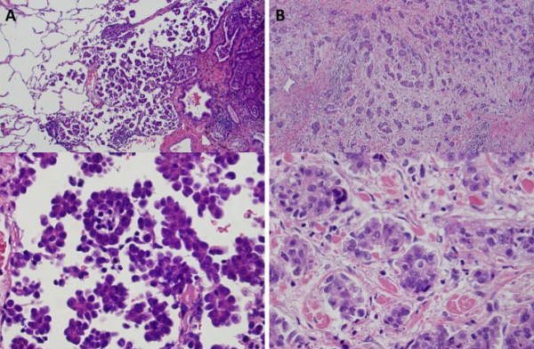
Pulmonary adenocarcinomas with a micropapillary component having small papillary tufts and lacking a central fibrovascular core are thought to result in poor prognosis. However, the component consists of tumor cells often floating within alveolar spaces (aerogenous micropapillary component [AMPC]) rather than invading fibrotic stroma observed in other organs like breast (stromal invasive micropapillary component [SMPC]). We previously observed cases of lung adenocarcinoma with predominant SMPC that was associated with micropapillary growth of tumors in fibrotic stroma observed in other organs. We evaluated the incidence and clinicopathological characteristics of SMPC in lung adenocarcinoma cases.
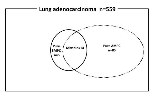
We investigated the clinicopathological characteristics and prognostic significance of SMPC in lung adenocarcinoma cases by reviewing 559 patients who had undergone surgical resection. We examined the SMPC by performing immunohistochemical analysis with 17 antibodies and by genetic analysis with epidermal growth factor receptor (EGFR) and KRAS mutations.
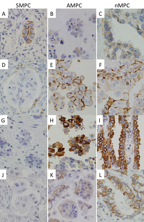
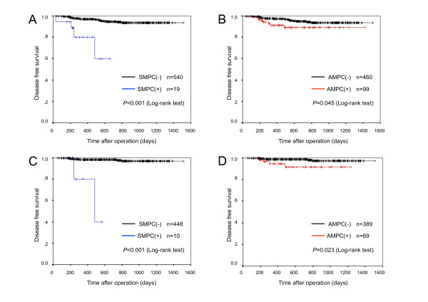
SMPC-positive (SMPC(+)) tumors were observed in 19 cases (3.4%). The presence of SMPC was significantly associated with tumor size, advanced-stage disease, lymph node metastasis, pleural invasion, lymphatic invasion, and vascular invasion. Patients with SMPC(+) tumors had significantly poorer outcomes than those with SMPC-negative tumors. Multivariate analysis revealed that SMPC was a significant independent prognostic factor of lung adenocarcinoma, especially for disease-free survival of pathological stage I patients (p = 0.035). SMPC showed significantly higher expression of E-cadherin and lower expression of CD44 than the corresponding expression levels shown by AMPC and showed lower surfactant apoprotein A and phospho-c-Met expression level than corresponding expression levels shown by tumor cell components without a micropapillary component. Fourteen cases with SMPC(+) tumors (74%) showed EGFR mutations, and none of them showed KRAS mutations.
SMPC(+) tumors are rare, but they may be associated with a poor prognosis and have different phenotypic and genotypic characteristics from those of AMPC(+) tumors.
具有小乳头状簇且缺乏中央纤维血管核心的微乳头状成分的肺腺癌被认为预后不良。然而,该成分由常常漂浮在肺泡空间中的肿瘤细胞组成(气源性微乳头状成分 [AMPC]),而不是像在其他器官(如乳腺)中观察到的侵袭性纤维间质的微乳头状成分(间质侵袭性微乳头状成分 [SMPC])。我们之前观察到肺腺癌病例中以 SMPC 为主,与其他器官中肿瘤在纤维间质中微乳头状生长有关。我们评估了肺腺癌病例中 SMPC 的发生率和临床病理特征。
我们通过复习 559 例接受手术切除的患者,研究了肺腺癌病例中 SMPC 的临床病理特征和预后意义。我们通过使用 17 种抗体进行免疫组织化学分析,并通过表皮生长因子受体(EGFR)和 KRAS 突变进行基因分析,来检测 SMPC。
在 19 例(3.4%)中观察到 SMPC 阳性(SMPC(+))肿瘤。SMPC 的存在与肿瘤大小、晚期疾病、淋巴结转移、胸膜侵犯、淋巴管侵犯和血管侵犯显著相关。SMPC(+)肿瘤患者的生存结局明显比 SMPC(-)肿瘤患者差。多变量分析显示,SMPC 是肺腺癌的一个显著独立预后因素,特别是对病理分期 I 期患者的无病生存(p=0.035)。SMPC 显示出比 AMPC 更高的 E-钙黏蛋白表达和更低的 CD44 表达,并且与无微乳头状成分的肿瘤细胞成分相比,SMPC 显示出更低的表面活性剂 A 蛋白和磷酸化 c-Met 表达水平。14 例 SMPC(+)肿瘤(74%)显示 EGFR 突变,且均无 KRAS 突变。
SMPC(+)肿瘤罕见,但它们可能与不良预后相关,并且与 AMPC(+)肿瘤具有不同的表型和基因型特征。