Department of Structural and Cellular Biology, Louisiana Cancer Research Consortium, New Orleans, LA 70112, USA.
BMC Cancer. 2012 Jan 10;12:10. doi: 10.1186/1471-2407-12-10.
The study of breast cancer metastasis depends on the use of established breast cancer cell lines that do not accurately represent the heterogeneity and complexity of human breast tumors. A tumor model was developed using primary breast tumor-initiating cells isolated from patient core biopsies that would more accurately reflect human breast cancer metastasis.
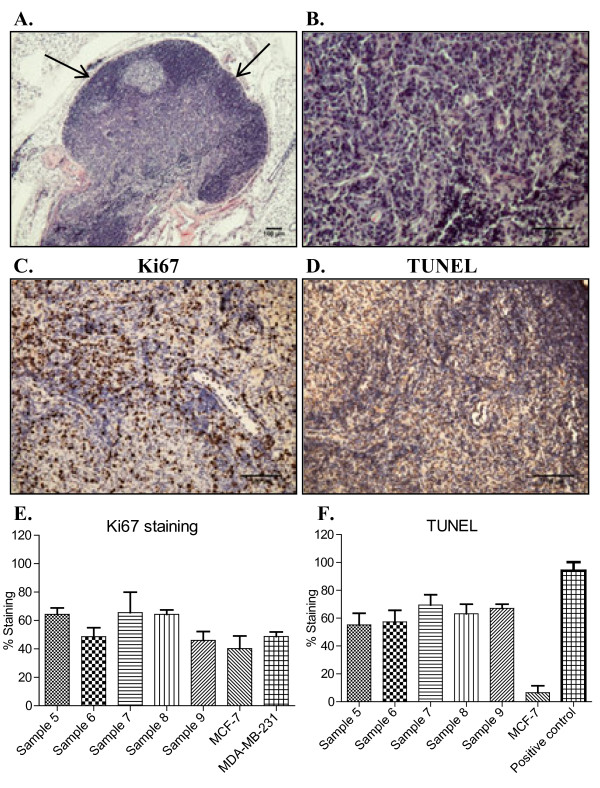
Tumorspheres were isolated under serum-free culture conditions from core biopsies collected from five patients with clinical diagnosis of invasive ductal carcinoma (IDC). Isolated tumorspheres were transplanted into the mammary fat pad of NUDE mice to establish tumorigenicity in vivo. Tumors and metastatic lesions were analyzed by hematoxylin and eosin (H+E) staining and immunohistochemistry (IHC).
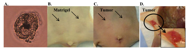
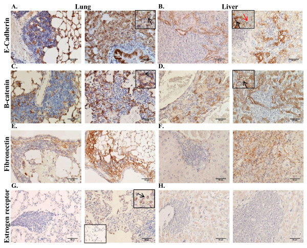
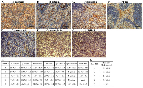
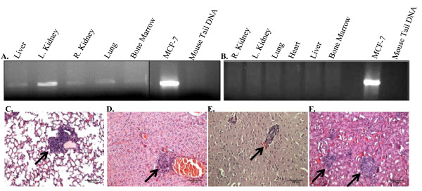
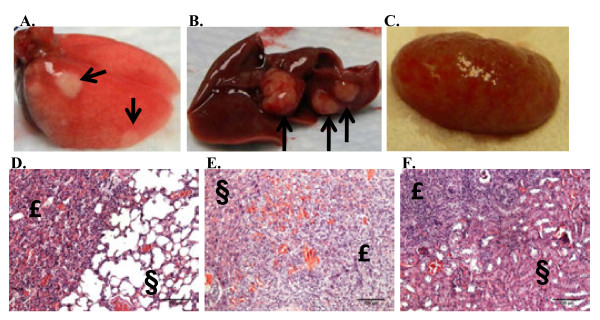
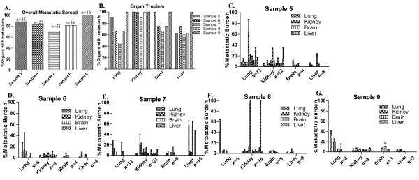
Tumorspheres were successfully isolated from all patient core biopsies, independent of the estrogen receptor α (ERα)/progesterone receptor (PR)/Her2/neu status or tumor grade. Each tumorsphere was estimated to contain 50-100 cells. Transplantation of 50 tumorspheres (1-5 × 10³ cells) in combination with Matrigel into the mammary fat pad of NUDE mice resulted in small, palpable tumors that were sustained up to 12 months post-injection. Tumors were serially transplanted three times by re-isolation of tumorspheres from the tumors and injection into the mammary fat pad of NUDE mice. At 3 months post-injection, micrometastases to the lung, liver, kidneys, brain and femur were detected by measuring content of human chromosome 17. Visible macrometastases were detected in the lung, liver and kidneys by 6 months post-injection. Primary tumors variably expressed cytokeratins, Her2/neu, cytoplasmic E-cadherin, nuclear β catenin and fibronectin but were negative for ERα and vimentin. In lung and liver metastases, variable redistribution of E-cadherin and β catenin to the membrane of tumor cells was observed. ERα was re-expressed in lung metastatic cells in two of five samples.
Tumorspheres isolated under defined culture conditions from patient core biopsies were tumorigenic when transplanted into the mammary fat pad of NUDE mice, and metastasized to multiple mouse organs. Micrometastases in mouse organs demonstrated a dormancy period prior to outgrowth of macrometastases. The development of macrometastases with organ-specific phenotypic distinctions provides a superior model for the investigation of organ-specific effects on metastatic cancer cell survival and growth.
乳腺癌转移的研究依赖于已建立的乳腺癌细胞系的使用,而这些细胞系并不能准确地代表人类乳腺癌肿瘤的异质性和复杂性。本研究通过使用从患者核心活检中分离得到的原发性乳腺癌起始细胞建立肿瘤模型,该模型更能准确反映人类乳腺癌转移。
在无血清培养条件下,从五名浸润性导管癌(IDC)临床诊断患者的核心活检中分离肿瘤球。将分离的肿瘤球移植到裸鼠的乳腺脂肪垫中,在体内建立致瘤性。通过苏木精和伊红(H+E)染色和免疫组织化学(IHC)分析肿瘤和转移病变。
所有患者的核心活检均成功分离出肿瘤球,与雌激素受体α(ERα)/孕激素受体(PR)/Her2/neu 状态或肿瘤分级无关。每个肿瘤球估计含有 50-100 个细胞。将 50 个肿瘤球(1-5×10³ 个细胞)与 Matrigel 一起移植到裸鼠的乳腺脂肪垫中,可形成小的、可触及的肿瘤,在注射后可持续长达 12 个月。通过从肿瘤中重新分离肿瘤球并注射到裸鼠的乳腺脂肪垫中,将肿瘤进行了三次连续的移植。在注射后 3 个月时,通过测量人 17 号染色体的含量,检测到肺、肝、肾、脑和股骨的微转移。在注射后 6 个月时,通过肉眼观察到肺、肝和肾中的大转移。原发肿瘤不同程度地表达细胞角蛋白、Her2/neu、细胞质 E-钙黏蛋白、核β连环蛋白和纤维连接蛋白,但 ERα 和波形蛋白呈阴性。在肺和肝转移中,观察到 E-钙黏蛋白和β连环蛋白在肿瘤细胞的膜上的分布发生了变化。在两个样本中的肺转移细胞中重新表达了 ERα。
从患者核心活检中在特定培养条件下分离出的肿瘤球在移植到裸鼠的乳腺脂肪垫中具有致瘤性,并转移到多个小鼠器官。在小鼠器官中的微转移在大转移的生长之前经历了休眠期。具有器官特异性表型差异的大转移的发展为研究转移癌细胞存活和生长的器官特异性影响提供了更好的模型。