Department of Biomedical Engineering, College of Medicine, Kyung Hee University, Seoul, Korea.
PLoS One. 2012;7(1):e30066. doi: 10.1371/journal.pone.0030066. Epub 2012 Jan 17.
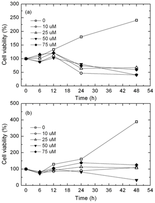
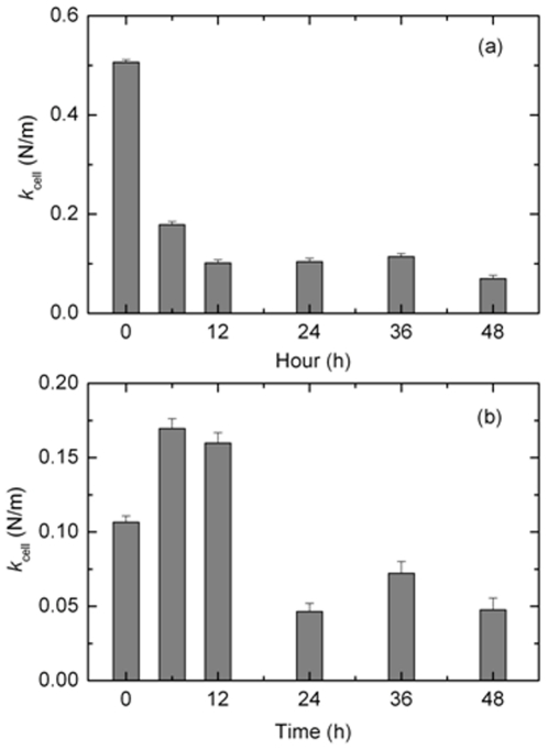
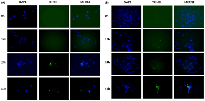
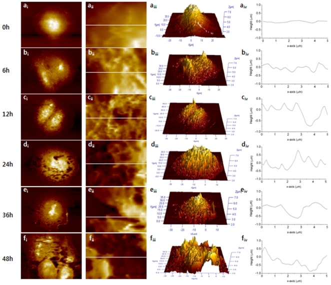
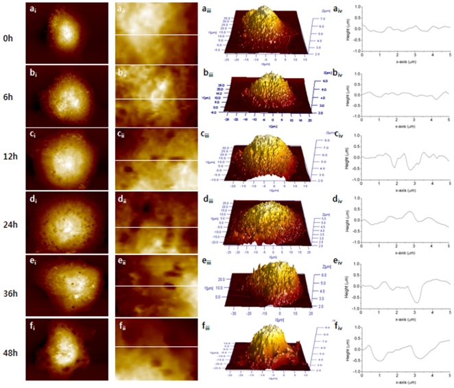
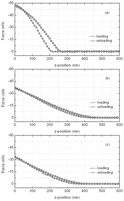
The apoptosis of cancer cells is associated with changes in the important cell properties including morphology, surface roughness and stiffness. Therefore, the changes in morphology and biophysical properties can be a good way of evaluating the anticancer activity of a drug. This study examined the effect of paclitaxel on the properties of Ishikawa and HeLa cells using atomic force microscopy (AFM), and the relationship between the changes in morphology and the biophysical properties and apoptosis was discussed. The viability and proliferation of the cells were analyzed using the methylthiazol tetrazolium (MTT) method and a TUNEL assay to confirm cellular apoptosis due to a paclitaxel treatment. AFM observations clearly showed the apoptotic morphological and biophysical changes in Ishikawa and HeLa cells. After the paclitaxel treatment, the cell membrane was torn and holed, the surface roughness was increased, and the stiffness was decreased. These changes were observed more apparently after a 24 h treatment and in Ishikawa cells compared to HeLa cells. The MTT and TUNEL assays results revealed the Ishikawa cells to be more sensitive to paclitaxel than HeLa cells and definite apoptosis occurred after a 24 h treatment. These results showed good agreement with the AFM results. Therefore, research on the morphological and biophysical changes by AFM in cancer cells will help to evaluate the anticancer activities of the drugs.
癌细胞的凋亡与包括形态、表面粗糙度和硬度在内的重要细胞特性的变化有关。因此,形态和生物物理特性的变化可以成为评估药物抗癌活性的一种很好的方法。本研究使用原子力显微镜(AFM)研究了紫杉醇对 Ishikawa 和 HeLa 细胞特性的影响,并讨论了形态变化与生物物理特性和细胞凋亡之间的关系。使用噻唑蓝(MTT)法和 TUNEL 检测法分析细胞活力和增殖,以确认紫杉醇处理导致的细胞凋亡。AFM 观察清楚地显示了 Ishikawa 和 HeLa 细胞的凋亡形态和生物物理变化。紫杉醇处理后,细胞膜撕裂并穿孔,表面粗糙度增加,硬度降低。与 HeLa 细胞相比,24 小时处理后,这些变化在 Ishikawa 细胞中更为明显。MTT 和 TUNEL 检测结果表明,Ishikawa 细胞对紫杉醇比 HeLa 细胞更为敏感,且在 24 小时处理后确实发生了凋亡。这些结果与 AFM 结果吻合较好。因此,通过 AFM 对癌细胞的形态和生物物理变化进行研究,将有助于评估药物的抗癌活性。