Institut de Génétique Humaine, Montpellier, France.
PLoS Genet. 2012 Jan;8(1):e1002465. doi: 10.1371/journal.pgen.1002465. Epub 2012 Jan 19.
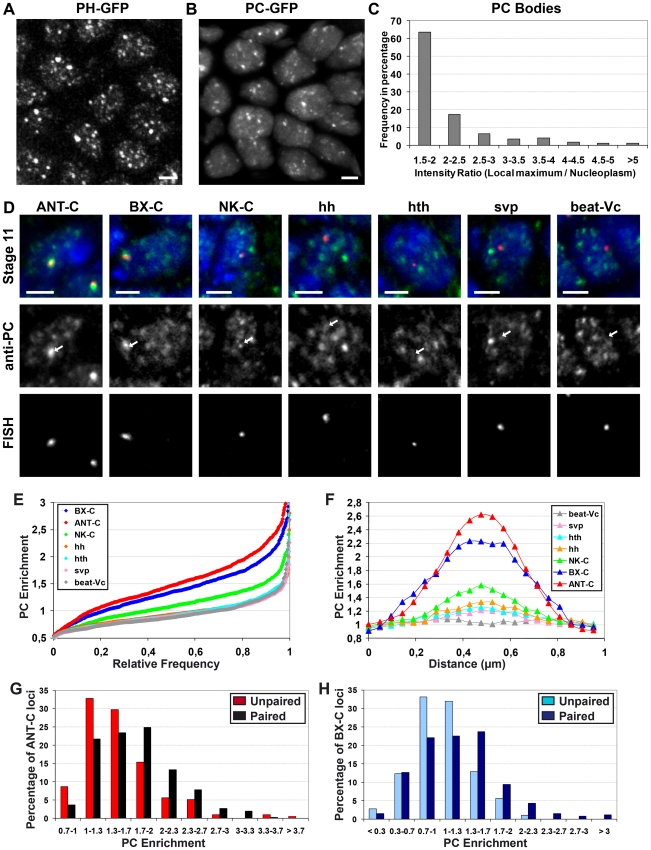
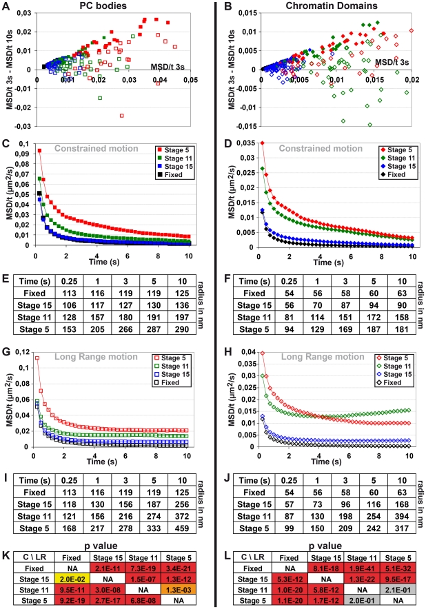
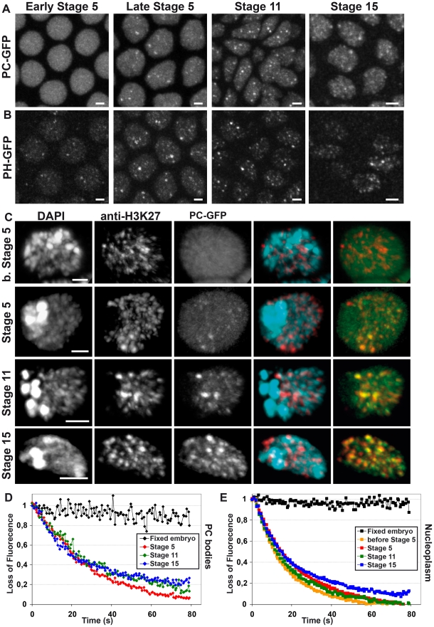
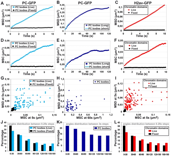
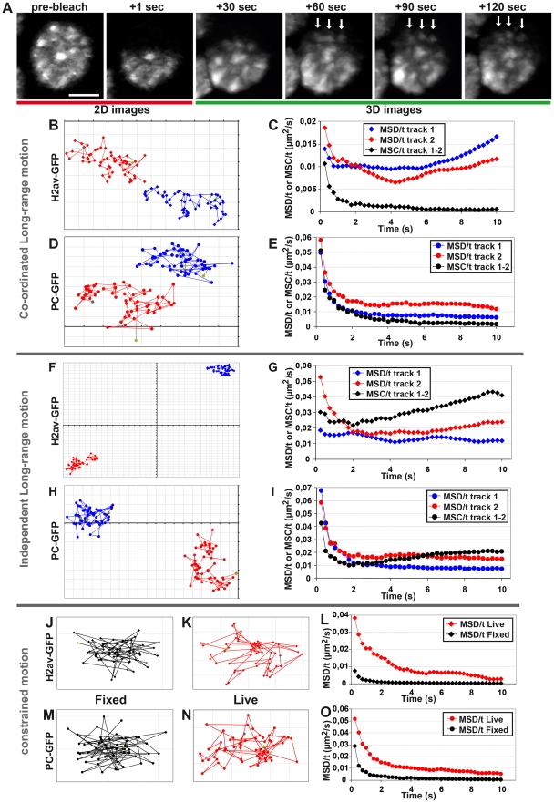
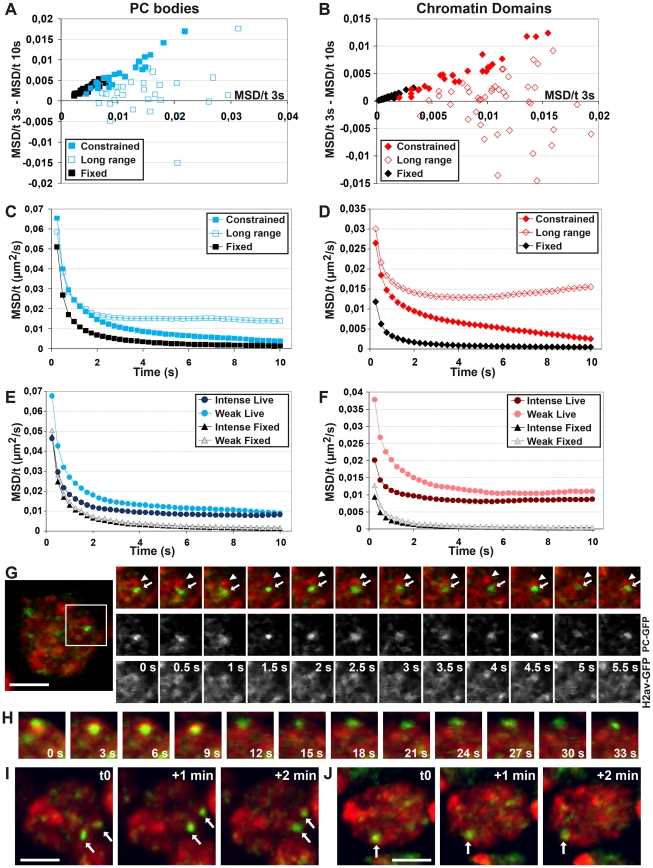
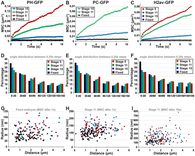
Polycomb group (PcG) proteins are conserved chromatin factors that maintain silencing of key developmental genes outside of their expression domains. Recent genome-wide analyses showed a Polycomb (PC) distribution with binding to discrete PcG response elements (PREs). Within the cell nucleus, PcG proteins localize in structures called PC bodies that contain PcG-silenced genes, and it has been recently shown that PREs form local and long-range spatial networks. Here, we studied the nuclear distribution of two PcG proteins, PC and Polyhomeotic (PH). Thanks to a combination of immunostaining, immuno-FISH, and live imaging of GFP fusion proteins, we could analyze the formation and the mobility of PC bodies during fly embryogenesis as well as compare their behavior to that of the condensed fraction of euchromatin. Immuno-FISH experiments show that PC bodies mainly correspond to 3D structural counterparts of the linear genomic domains identified in genome-wide studies. During early embryogenesis, PC and PH progressively accumulate within PC bodies, which form nuclear structures localized on distinct euchromatin domains containing histone H3 tri-methylated on K27. Time-lapse analysis indicates that two types of motion influence the displacement of PC bodies and chromatin domains containing H2Av-GFP. First, chromatin domains and PC bodies coordinately undergo long-range motions that may correspond to the movement of whole chromosome territories. Second, each PC body and chromatin domain has its own fast and highly constrained motion. In this motion regime, PC bodies move within volumes slightly larger than those of condensed chromatin domains. Moreover, both types of domains move within volumes much smaller than chromosome territories, strongly restricting their possibility of interaction with other nuclear structures. The fast motion of PC bodies and chromatin domains observed during early embryogenesis strongly decreases in late developmental stages, indicating a possible contribution of chromatin dynamics in the maintenance of stable gene silencing.
多梳组 (PcG) 蛋白是保守的染色质因子,可在外显子表达域之外维持关键发育基因的沉默。最近的全基因组分析显示,多梳(PC)蛋白与离散的 PcG 反应元件 (PRE) 结合分布。在细胞核内,PcG 蛋白定位于称为 PC 体的结构中,这些结构包含 PcG 沉默的基因,最近有研究表明 PRE 形成局部和长程空间网络。在这里,我们研究了两种 PcG 蛋白 PC 和 Polyhomeotic (PH) 的核分布。得益于免疫染色、免疫荧光原位杂交(immuno-FISH)和 GFP 融合蛋白的活细胞成像的组合,我们可以在果蝇胚胎发生过程中分析 PC 体的形成和流动性,并将其与常染色质浓缩部分的行为进行比较。免疫荧光原位杂交实验表明,PC 体主要对应于全基因组研究中线性基因组域的 3D 结构对应物。在早期胚胎发生过程中,PC 和 PH 逐渐积累在 PC 体中,PC 体形成定位于含有 H3 第 27 位三甲基化的特定常染色质域的核结构。时程分析表明,两种类型的运动影响 PC 体和含有 H2Av-GFP 的染色质域的位移。首先,染色质域和 PC 体协调地进行长程运动,这可能对应于整个染色体区域的运动。其次,每个 PC 体和染色质域都有自己的快速且高度受限的运动。在这种运动状态下,PC 体在体积稍大于浓缩染色质域的体积内移动。此外,这两种类型的域都在体积明显小于染色体区域的体积内移动,强烈限制了它们与其他核结构相互作用的可能性。在胚胎发生早期观察到的 PC 体和染色质域的快速运动在发育后期阶段强烈减少,表明染色质动力学可能有助于维持稳定的基因沉默。