Center for Vision Research, Department of Neuroscience, State University of New York Upstate Medical University, Syracuse, New York, United States of America.
PLoS One. 2012;7(1):e30101. doi: 10.1371/journal.pone.0030101. Epub 2012 Jan 19.
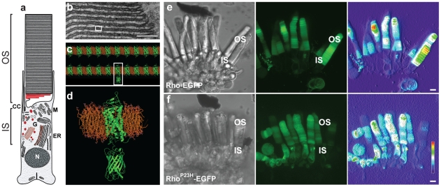
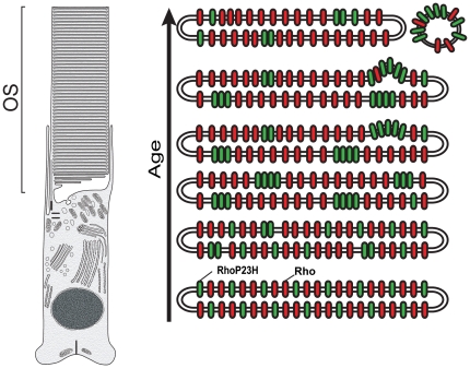
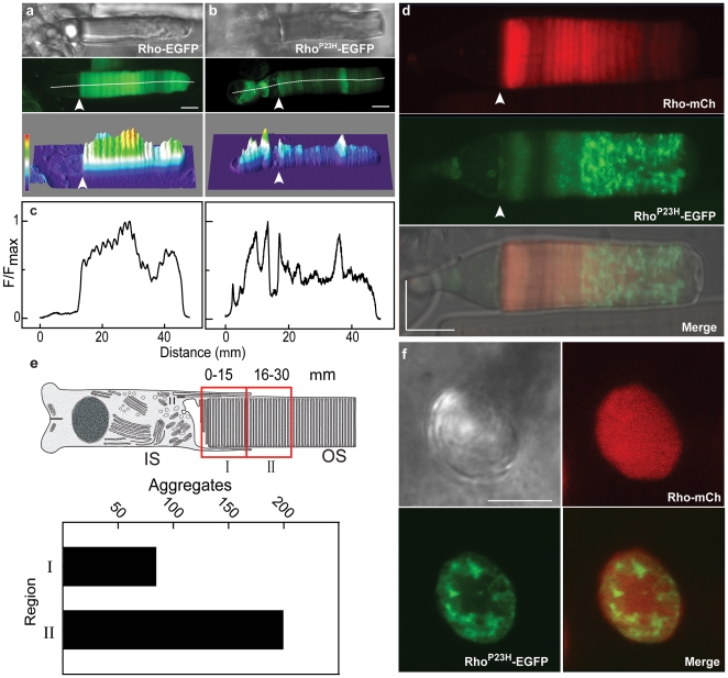
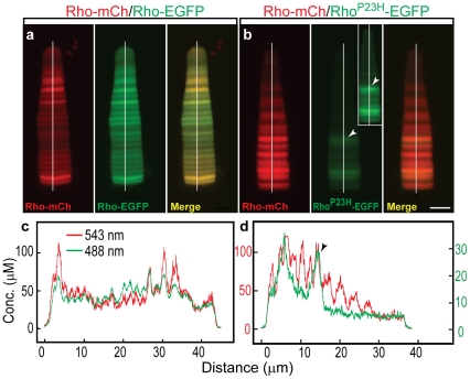
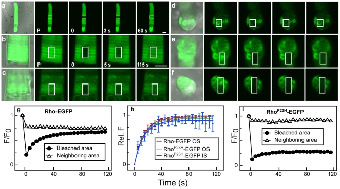
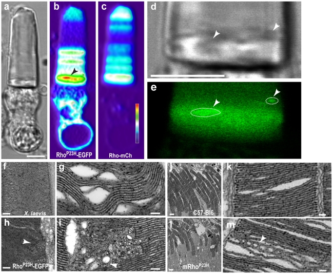
Mutations in rhodopsin cause retinitis pigmentosa in humans and retinal degeneration in a multitude of other animals. We utilized high-resolution live imaging of the large rod photoreceptors from transgenic frogs (Xenopus) to compare the properties of fluorescently tagged rhodopsin, Rho-EGFP, and Rho(P23H)-EGFP. The mutant was abnormally distributed both in the inner and outer segments (OS), accumulating in the OS to a concentration of ∼0.1% compared to endogenous opsin. Rho(P23H)-EGFP formed dense fluorescent foci, with concentrations of mutant protein up to ten times higher than other regions. Wild-type transgenic Rho-EGFP did not concentrate in OS foci when co-expressed in the same rod with Rho(P23H)-EGFP. Outer segment regions containing fluorescent foci were refractory to fluorescence recovery after photobleaching, while foci in the inner segment exhibited recovery kinetics similar to OS regions without foci and Rho-EGFP. The Rho(P23H)-EGFP foci were often in older, more distal OS disks. Electron micrographs of OS revealed abnormal disk membranes, with the regular disk bilayers broken into vesiculotubular structures. Furthermore, we observed similar OS disturbances in transgenic mice expressing Rho(P23H), suggesting such structures are a general consequence of mutant expression. Together these results show that mutant opsin disrupts OS disks, destabilizing the outer segment possibly via the formation of aggregates. This may render rods susceptible to mechanical injury or compromise OS function, contributing to photoreceptor loss.
视紫红质突变会导致人类的色素性视网膜炎和多种其他动物的视网膜变性。我们利用转基因青蛙(非洲爪蟾)大杆状光感受器的高分辨率实时成像,比较了荧光标记的视紫红质、Rho-EGFP 和 Rho(P23H)-EGFP 的特性。该突变体在内节和外节(OS)中分布异常,与内源性视蛋白相比,在外节中积累到约 0.1%的浓度。Rho(P23H)-EGFP 形成密集的荧光焦点,突变蛋白的浓度比其他区域高十倍以上。当与 Rho(P23H)-EGFP 在同一根杆状细胞中共同表达时,野生型转基因 Rho-EGFP 不会在外节焦点中浓缩。含有荧光焦点的外节区域对光漂白后的荧光恢复具有抗性,而内节中的焦点表现出与无焦点和 Rho-EGFP 的 OS 区域相似的恢复动力学。Rho(P23H)-EGFP 焦点通常位于较老、更远的 OS 盘上。OS 的电子显微镜照片显示出异常的盘膜,正常的盘双层被破坏成囊泡管状结构。此外,我们在表达 Rho(P23H)的转基因小鼠中观察到类似的 OS 紊乱,表明这些结构是突变体表达的一般后果。这些结果表明,突变的视蛋白会破坏 OS 盘,通过形成聚集体使外节不稳定,这可能使杆状细胞容易受到机械损伤或损害 OS 功能,从而导致光感受器丧失。